A common practitioner mistake: attributing post-viral cognitive symptoms to lingering viral load when the actual driver is ongoing neuroinflammation. The virus may be gone. The immune response it triggered may not be. Dr. Datis Kharrazian's clinical training at the Kharrazian Institute addresses this distinction directly, because missing it leads to treatment strategies that target the wrong mechanism entirely.
Viral infections neuroinflammation symptoms are not the same as active infection symptoms. The clinical picture looks similar on the surface, which is why so many patients remain unresolved.
How Viral Infections Trigger Brain Inflammation
When a virus enters the body, the immune system mounts a response that extends well beyond the peripheral tissues. Neuroinflammation research demonstrates that viral pathogens can activate the brain's resident immune cells, the microglia and astrocytes, either through direct CNS invasion or through peripheral immune signaling that crosses the blood-brain barrier via cytokine cascades.
Once glial cells are activated, they release pro-inflammatory cytokines including IL-1β, IL-6, and TNF-α. These cytokines suppress neuronal ATP production, interfere with neurotransmitter synthesis, and degrade the myelin that facilitates axonal conduction. The result is a brain that is metabolically impaired and structurally stressed, regardless of whether the virus is still replicating.
The critical teaching point in Dr. Kharrazian's coursework: the brain does not have a simple off-switch for this response. Glial activation, once initiated, can persist for months or years after the initial infection has cleared. This is not metaphor. It is measurable neuroimmune physiology.
Why Specific Brain Regions Produce Specific Symptoms
Neuroinflammation is not diffuse by nature. Regional blood flow patterns, local metabolic demands, and pre-existing vulnerabilities determine which areas of the brain sustain the most significant inflammatory burden. This regional specificity is what produces the symptom patterns practitioners see clinically.
Inflammation concentrated in the left premotor cortex, for instance, manifests as word-retrieval difficulty and disrupted speech production. Patients describe knowing what they want to say but being unable to access the word quickly. This is not evidence that the virus persists in brain tissue. It is evidence of focal neuroinflammation impairing a specific functional region. The distinction matters because the clinical strategy changes entirely depending on which mechanism is driving the presentation.
Prefrontal cortex involvement produces executive dysfunction, poor working memory, and difficulty sequencing tasks. Cerebellar inflammation alters coordination and gait. Limbic involvement drives mood dysregulation and anxiety disproportionate to the patient's life circumstances. These are not vague post-viral complaints. They are neuroanatomically logical consequences of regional immune activation, and research in neuroimmunology has documented them in detail. Dr. Kharrazian's clinical training teaches practitioners to map symptoms back to these regions systematically, rather than treating the full constellation as a single undifferentiated problem.
The Metabolic Collapse Behind Infection-Induced Brain Fog
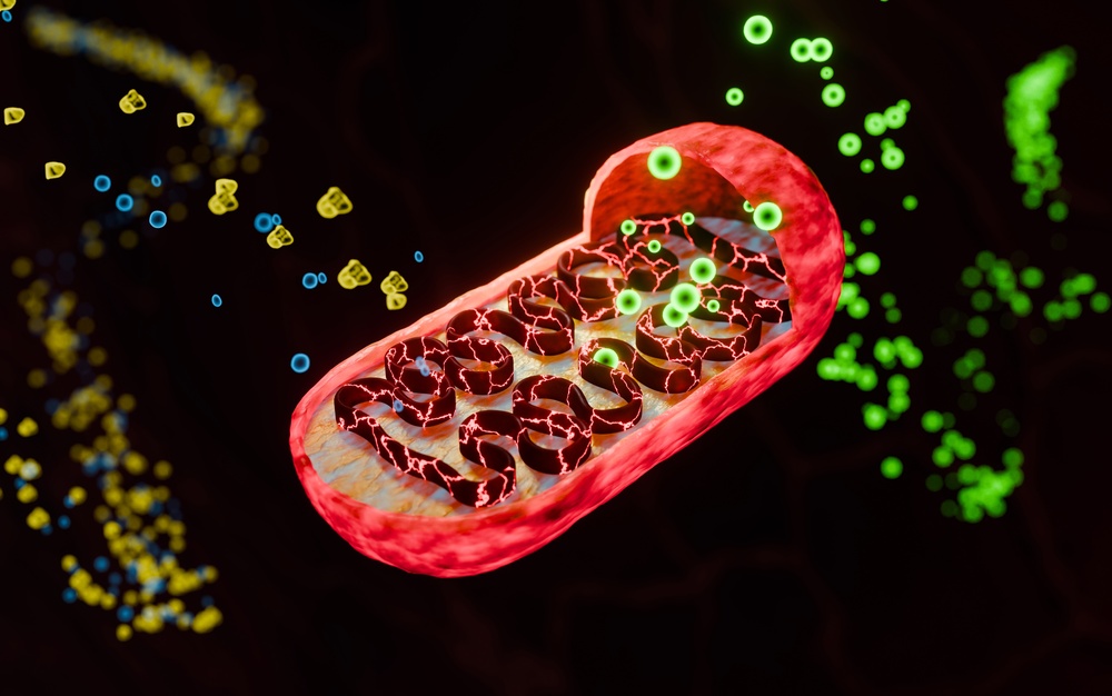
Brain fog is a metabolic event before it is a cognitive one. Cytokine-driven neuroinflammation suppresses mitochondrial function in neurons, reducing ATP output in brain tissue. When neuronal ATP drops, the sodium-potassium pump that maintains membrane excitability slows. Neurons fire less efficiently. Synaptic transmission degrades. The patient experiences this as cognitive sluggishness, reduced processing speed, and the inability to sustain mental effort for more than short intervals.
Infection-induced brain fog has a specific signature: it is disproportionately worse after cognitive exertion. Patients often describe a functional "ceiling" they can work up to before a sharp drop occurs. This is post-exertional neural fatigue driven by metabolic depletion, not psychological burnout. The research synthesized in Dr. Kharrazian's coursework connects this pattern directly to glial-mediated mitochondrial suppression.
The clinical implication is that pushing these patients through high-demand cognitive work without addressing the underlying neuroinflammatory and metabolic state consistently produces setbacks. The brain's energy substrate is insufficient for the load being placed on it.
Chronic Viral Infections and Sustained Neuroinflammatory States
Acute infections resolve. Chronic infections — Epstein-Barr, cytomegalovirus, HHV-6, and others with well-documented latency mechanisms — do not fully clear. They establish reservoirs in immune and tissue cells and reactivate under conditions of immune suppression, physiological stress, or nutrient depletion. Each reactivation event re-triggers glial activation and re-elevates the neuroinflammatory baseline.
The clinical pattern in these patients is cyclical deterioration. They stabilize partially, then relapse when stressed, sleep-deprived, or physically ill. Practitioners who are not tracking the viral reactivation component often interpret these relapses as evidence that a different condition is progressing, rather than recognizing the immune-brain cycle for what it is.
Research in immunology and neuroimmunology documents that sustained immune activation from chronic viral infections also depletes regulatory T-cell populations, which are responsible for suppressing excessive immune responses. When regulatory function degrades, the immune system loses its capacity to self-limit, and the neuroinflammatory state becomes self-reinforcing independent of the viral trigger itself.
Molecular Mimicry and Autoimmune Neurological Damage
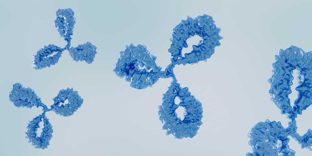
Persistent viral infections introduce a second mechanism of neurological damage: autoimmunity through molecular mimicry. Certain viral proteins share structural sequences with human neuronal proteins. When the immune system generates antibodies against viral epitopes, those antibodies can cross-react with host tissue. The immune system is not malfunctioning. It is responding accurately to a pathogen but generating collateral damage against self-tissue in the process.
Research in autoimmunity has identified molecular mimicry as a factor in post-infectious neurological syndromes, including conditions affecting myelin integrity, cerebellar function, and ganglionic nerve tissue. Dr. Kharrazian's clinical training addresses how to assess for this pattern in patients whose neurological symptoms persist and progress long after an apparent infectious resolution.
The clinical picture in these cases diverges from straightforward post-viral neuroinflammation in one important way: the symptoms do not stabilize when the infection is managed. They continue to progress because the driver has shifted from the virus to the immune system's now-autonomous attack on neural tissue. Identifying that shift determines the entire clinical strategy.
Glial Priming and the Risk of Disproportionate Reactions
Each episode of glial activation lowers the threshold for the next one. This phenomenon, glial priming, is documented in neuroinflammation research and represents one of the most clinically significant concepts for practitioners treating patients with repeated infections or long-duration post-viral symptoms.
A primed microglial population responds to subsequent immune challenges, whether from a new infection, a metabolic stressor, or a toxin exposure, with an exaggerated inflammatory response. The patient appears to react to triggers that would have minimal impact on someone without prior glial activation history. Practitioners unfamiliar with this mechanism often interpret these exaggerated responses as evidence of a new and separate condition. In many cases, they are a predictable consequence of accumulated neuroinflammatory history.
Dr. Kharrazian's coursework teaches practitioners to take a thorough viral history not because prior infections are necessarily the current problem, but because they establish the neuroimmune context in which the current problem is occurring. That context determines how aggressively the patient's brain will respond to even modest new triggers, and it informs realistic expectations about the pace of recovery.
Clinical Strategies for Addressing Viral Neuroinflammation
Treating neuroinflammation from viral infections requires working across multiple mechanisms simultaneously. Addressing only one leaves the others active.
- Assess immune activation state: Cytokine panels, complement markers, and viral reactivation antibody titers provide data on whether the immune-brain axis is currently being driven by active viral reactivation or by autonomous post-infectious immune dysregulation.
- Support microglial regulation: Research in neuroinflammation identifies several nutritional and botanical compounds with documented effects on microglial activation, including palmitoylethanolamide, luteolin, and specific polyphenols. The clinical application of these compounds is covered in depth in Dr. Kharrazian's coursework.
- Address mitochondrial function: Neuronal ATP deficits do not self-correct while inflammation remains elevated, but supporting mitochondrial substrate availability can reduce the functional impact while broader resolution occurs.
- Evaluate for autoimmune crossover: Patients with progressive rather than stable post-viral neurological symptoms warrant assessment for neural autoantibodies. The presence of autoimmunity changes both the target of treatment and the expected timeline.
- Manage reactivation triggers: Sleep deprivation, psychological stress, nutrient depletion, and secondary infections are the most common reactivation triggers for latent viral pathogens. Systematic identification and reduction of these factors is not supportive care. It is a core component of the clinical strategy.
Key Takeaways
- Viral infections neuroinflammation symptoms can persist and progress long after active infection has resolved, driven by ongoing glial activation rather than persistent viral load.
- Neuroinflammation is regionally specific. The symptom pattern reflects which brain areas are carrying the highest inflammatory burden.
- Infection-induced brain fog is a metabolic event driven by cytokine suppression of neuronal ATP production, not a psychological response to illness.
- Chronic latent viral infections create cyclical neuroinflammatory reactivation patterns that require tracking viral immune status, not just current symptoms.
- Molecular mimicry can shift the driver of neurological damage from the virus to autonomous autoimmune activity, changing the clinical strategy entirely.
Frequently Asked Questions
Research in neuroimmunology confirms that glial activation initiated by a viral infection can persist for extended periods. Chronic latent infections, glial priming from repeated exposures, and autoimmune crossover are all mechanisms that sustain neuroinflammation well beyond the acute infectious episode.
Post-viral brain fog is primarily driven by cytokine-mediated suppression of neuronal mitochondrial function. When brain ATP drops, neuronal firing efficiency drops with it. The result is reduced processing speed, impaired working memory, and post-exertional cognitive fatigue that worsens with mental effort.
Viral infections trigger glial cell activation in the brain, releasing pro-inflammatory cytokines that interfere with neurotransmitter synthesis, reduce myelin integrity, and suppress neuronal energy production. The severity and location of this neuroinflammation determine the specific cognitive dysfunction a patient experiences.
Molecular mimicry is a well-documented mechanism in which viral proteins share structural sequences with human neural proteins. Immune responses targeting the virus can cross-react with host neuronal tissue, initiating autoimmune processes that continue progressing even after the original infection is controlled.
Assessment should include viral reactivation antibody titers, markers of immune activation, neurological autoantibody panels, and a detailed history of prior infections. Together these distinguish active viral reactivation from autonomous post-infectious autoimmunity, which require different clinical strategies.
About the Author
Dr. Datis Kharrazian, PhD, DHSc, DC, MS, MMSc, FACN is a Harvard Medical School research fellow and researcher at Massachusetts General Hospital Department of Neurology specializing in autoimmunity and neuroimmunology. He serves as Associate Clinical Professor at Loma Linda University School of Medicine and is the author of Why Do I Still Have Thyroid Symptoms When My Lab Tests Are Normal and Why Isn't My Brain Working. He is a Fellow of the American College of Nutrition, Diplomate of the Board of Nutrition Specialists, a member of the American Association of Immunologists, and a Fellow of the Royal Society of Medicine (UK). The Kharrazian Institute serves more than 5,000 physicians and healthcare providers worldwide.
Practitioners seeking to implement these clinical strategies can explore Dr. Kharrazian's coursework on neuroinflammation and immune-brain axis dysfunction at the Kharrazian Institute.