Most practitioners ordering longevity panels are measuring the wrong things. Telomere length, inflammatory markers, and lipid oxidation panels tell you something about aging — but they don't tell you why the cell is failing. Mitochondrial health longevity testing gets closer to the actual mechanism. The Kharrazian Institute's advanced longevity training teaches practitioners how to assess mitochondrial function systematically, because the clinical presentations of mitochondrial decline — fatigue that doesn't respond to sleep, cognitive slowing, emotional fragility, impaired exercise recovery — are frequently misattributed to other causes while the cell's energy infrastructure quietly deteriorates.
The reason this matters clinically is direct: every organ system runs on ATP. When ATP production drops, the highest-demand tissues fail first. That's the brain, the heart, and the skeletal muscle — precisely the organ systems most associated with aging outcomes.
Why Mitochondrial Dysfunction Is Central to the Aging Process
Aging research consistently identifies mitochondrial decline as one of the primary biological drivers of cellular senescence. This isn't a functional medicine-specific claim — it's a finding that has emerged from decades of molecular biology, replicated across model organisms and human tissue studies.
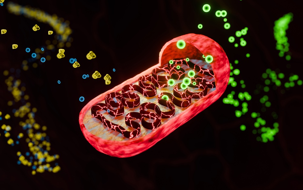
Two processes govern mitochondrial health over a lifetime: biogenesis (the creation of new mitochondria) and mitophagy (the selective clearance of damaged ones). Both decline with age. As biogenesis slows, the cell has fewer mitochondria available for energy production. As mitophagy becomes less efficient, damaged mitochondria accumulate instead of being cleared. The result is a population of dysfunctional organelles generating reactive oxygen species without producing adequate ATP — oxidative stress rising while energy output falls.
Mitochondrial fusion and fission add another layer. Fusion allows healthy mitochondria to share components and compensate for localized damage. Fission isolates damaged segments so they can be cleared via mitophagy. When this balance shifts — typically toward excess fission in aging tissue — mitochondrial fragmentation accelerates and bioenergetic capacity collapses. Research in mitochondrial biology has mapped this process across aging tissue with considerable precision. Dr. Kharrazian's clinical training teaches practitioners to recognize the functional downstream effects of this collapse and to connect those clinical presentations to the underlying biology.
What Clinical Markers Actually Reflect Mitochondrial Status
No single lab value measures mitochondrial function directly in a standard clinical panel. What practitioners can do is build an indirect picture from multiple converging markers. Dr. Kharrazian's coursework at the Kharrazian Institute addresses this gap by teaching a structured approach to marker selection and interpretation.
Organic acids testing is among the most clinically useful tools available. Elevated markers such as citric acid cycle intermediates (particularly succinate, fumarate, and malate) or markers of impaired beta-oxidation indicate mitochondrial inefficiency at specific metabolic steps. Lactate-to-pyruvate ratios can signal impaired electron transport chain activity. These aren't obscure findings — they're interpretable patterns that point to where in the mitochondrial pathway the breakdown is occurring.
Additional markers worth integrating into a mitochondrial assessment include:
- CoQ10 levels: Coenzyme Q10 is essential for electron transfer within the mitochondrial membrane. Depletion — particularly common in patients on statin therapy — directly impairs ATP synthesis.
- Plasma or urine carnitine fractions: Carnitine is required to transport long-chain fatty acids into the mitochondrial matrix for beta-oxidation. Low free carnitine or an elevated acylcarnitine-to-free-carnitine ratio reflects impaired fatty acid utilization.
- Glutathione status: Mitochondria are the primary site of reactive oxygen species generation. Glutathione is the primary intracellular antioxidant defense. Depletion reflects oxidative burden beyond what the cell can manage.
- Fasting insulin and glucose: Insulin resistance at the cellular level impairs mitochondrial glucose utilization. Chronically elevated insulin is one of the most modifiable drivers of mitochondrial inefficiency.
- NAD+ precursor status: NAD+ is a critical cofactor in the electron transport chain. Its decline with age is one of the most researched contributors to mitochondrial aging. Functional markers of NAD+ status can be inferred through NMN/NR metabolites on organic acids panels.
The clinical value is in the pattern, not any single value. A patient with low CoQ10, elevated lactate-to-pyruvate ratio, and impaired fasting glucose has a convergent picture of mitochondrial compromise — regardless of what their standard metabolic panel shows.
How Autophagy and Mitophagy Connect to Longevity Assessment
Autophagy is the cell's internal recycling system. Mitophagy is its mitochondria-specific branch. When either process is impaired, cellular debris accumulates — damaged proteins, dysfunctional organelles, fragmented mitochondrial membranes. That accumulation is a core feature of aged tissue.
Research in the longevity field has identified autophagy impairment as a convergence point between mitochondrial dysfunction, neurodegeneration, and chronic disease susceptibility. This isn't a marginal finding — it's why caloric restriction, intermittent fasting, and certain nutraceuticals (rapamycin analogues in research settings, berberine and spermidine in clinical application) have attracted serious scientific interest as longevity interventions. They share a common mechanism: autophagy induction.
From a clinical testing standpoint, autophagy activity isn't directly measurable in routine practice. What practitioners can assess are the conditions that suppress it. Chronically elevated insulin suppresses autophagy. Persistent mTOR activation from excess caloric load does the same. Sleep deprivation impairs the circadian regulation of autophagic flux. These are all clinically addressable. The Kharrazian Institute's training on longevity emphasizes that mitochondrial health longevity testing isn't only about measuring markers — it's about identifying the suppressors of cellular repair that are modifiable through clinical intervention.
Exercise, Sleep, and Metabolic Inputs as Testable Variables
Mitochondrial biogenesis is not exclusively a pharmaceutical target. It is a physiological process that responds to specific lifestyle inputs with measurable effects. PGC-1α, the master regulator of mitochondrial biogenesis, is activated by endurance exercise, cold exposure, caloric restriction, and certain polyphenols. The clinical implication: a patient's exercise behavior, sleep architecture, and metabolic status are directly relevant to their mitochondrial function — not as background history, but as active variables that need to be assessed.
A sedentary patient with disrupted sleep and chronically elevated blood glucose has a predictable mitochondrial phenotype before a single lab is ordered. The labs confirm and quantify — they don't replace — clinical pattern recognition.
Sleep specifically deserves attention here. Autophagy in the brain follows a circadian rhythm that peaks during deep sleep. Impaired sleep doesn't just impair cognitive recovery; it impairs the clearance of the damaged mitochondrial components that accumulate during waking neurological activity. Research in sleep biology and neurodegeneration has increasingly linked chronic sleep disruption to mitochondrial fragmentation in neural tissue. Asking a longevity patient about their sleep is not a soft clinical question. It's a direct inquiry into their mitophagy rate.
Nutraceutical Support: Mechanism-Based, Not Symptomatic
The research synthesized in Dr. Kharrazian's coursework gives practitioners a mechanism-based rationale for nutraceutical selection in mitochondrial support — which is different from symptom-based supplementation. Fatigue does not equal CoQ10 deficiency. The clinical decision requires knowing where in the mitochondrial pathway the dysfunction lies.
Patients with impaired electron transport chain function require different support than patients with impaired beta-oxidation. Patients with elevated oxidative stress markers need antioxidant support upstream of mitochondrial function — not simply more ATP precursors. Patients with NAD+ depletion respond to different interventions than those with carnitine insufficiency. The testing informs the protocol, and the protocol targets the mechanism rather than the symptom cluster.
This is the clinical application Dr. Kharrazian emphasizes: that nutraceuticals with genuine mitochondrial research behind them — CoQ10, alpha-lipoic acid, acetyl-L-carnitine, NAD+ precursors, PQQ — are not interchangeable. Their appropriate use depends on correctly identifying which aspect of mitochondrial function is compromised in the individual patient.
Building a Mitochondrial Assessment Into a Longevity Protocol
A structured mitochondrial assessment for longevity practice incorporates four dimensions: lab markers, lifestyle inputs, organ system demand (which tissues are highest-demand for that patient), and symptom pattern. No single dimension is sufficient alone.
A patient presenting with cognitive slowing, morning fatigue despite adequate sleep, and flat affect who also shows elevated organic acid markers for citric acid cycle dysfunction is a very different clinical picture than a patient with the same symptoms and normal organic acids but chronically elevated fasting insulin. The interventions diverge significantly. The former may require direct mitochondrial cofactor support; the latter requires metabolic stabilization before mitochondrial support is even fully addressable.
Dr. Kharrazian's clinical training at the Kharrazian Institute teaches practitioners to hold this level of specificity — not to reach for a mitochondrial support protocol as a default response to fatigue, but to identify which patients have which type of mitochondrial compromise and to design interventions that match the mechanism.
Longevity medicine that doesn't assess mitochondrial function is addressing the margins of aging rather than the core. The cell's energy infrastructure determines every downstream process that practitioners are trying to optimize.
Key Takeaways
- Mitochondrial dysfunction — driven by declining biogenesis, impaired mitophagy, and disrupted fusion/fission balance — is a primary mechanism of cellular aging, not a secondary consequence of it.
- No single lab value measures mitochondrial function directly; organic acids, CoQ10, carnitine fractions, glutathione, and fasting insulin form a converging clinical picture when interpreted together.
- Autophagy suppression (from elevated insulin, poor sleep, or excess caloric load) is a modifiable clinical variable — not just a research concept.
- Nutraceutical selection for mitochondrial support should follow mechanism identification, not symptom pattern alone.
- Exercise behavior, sleep quality, and metabolic status are active mitochondrial variables — clinically assessable before a single lab is ordered.
Frequently Asked Questions
No single test measures mitochondrial function in standard clinical practice. Organic acids testing provides the most detailed picture of mitochondrial pathway efficiency. Combined with CoQ10 levels, carnitine fractions, glutathione status, and fasting insulin, practitioners can build a reliable indirect assessment of where mitochondrial function is compromised.
The most common presentations are fatigue unresponsive to adequate sleep, impaired exercise recovery, cognitive slowing, emotional fragility, and reduced stress tolerance. These symptoms reflect inadequate ATP production in high-demand tissues — brain, heart, and skeletal muscle — and are frequently misattributed to other causes.
Endurance exercise is the most reliably documented activator of PGC-1α, the primary regulator of mitochondrial biogenesis. Caloric restriction, intermittent fasting, stable blood glucose, adequate sleep, and certain polyphenols also support biogenesis and autophagic clearance of damaged mitochondria.
Yes. Mitochondrial decline precedes symptomatic presentation by years. In longevity-focused practice, mitochondrial assessment is relevant as a preventive measure — identifying subclinical dysfunction in organ systems like the brain or cardiovascular tissue before functional impairment becomes the chief complaint.
Insulin resistance impairs the cell's ability to utilize glucose for mitochondrial ATP production and suppresses autophagy. Chronically elevated insulin is one of the most common and modifiable drivers of mitochondrial dysfunction — making metabolic stabilization a prerequisite for effective mitochondrial support in many patients.
About the Author
Dr. Datis Kharrazian, PhD, DHSc, DC, MS, MMSc, FACN is a Harvard Medical School research fellow and researcher at Massachusetts General Hospital's Department of Neurology, specializing in autoimmunity and neuroimmunology. He serves as Associate Clinical Professor at Loma Linda University School of Medicine and is the author of Why Do I Still Have Thyroid Symptoms When My Lab Tests Are Normal and Why Isn't My Brain Working. He is a Fellow of the American College of Nutrition, a Diplomate of the Board of Nutrition Specialists, a member of the American Association of Immunologists, and a Fellow of the Royal Society of Medicine (UK). The Kharrazian Institute serves more than 5,000 physicians and healthcare providers worldwide.
Train in Advanced Longevity Medicine
The Kharrazian Institute offers clinical training in mitochondrial assessment and longevity protocols for licensed healthcare providers. The coursework covers mechanism-based testing interpretation, personalized intervention strategies, and clinical application for complex chronic patients. Details are available at the Kharrazian Institute website.