Initiating a detox protocol on the wrong patient is one of the most consequential clinical mistakes a practitioner can make. Not because detoxification is inherently dangerous, but because the practitioners most likely to pursue it are also the most likely to attract complex, chronic patients whose systems cannot sustain the physiological demands it creates. Dr. Datis Kharrazian and the Kharrazian Institute have addressed this problem directly in clinical training, and the answer is not a checklist — it is a thought process built on recognizing specific clinical patterns before the protocol begins.
Heavy metal chelation and aggressive detox protocols mobilize stored toxins and move them through the body's clearance pathways. In a patient with intact barriers, competent liver and kidney function, and a regulated immune system, that process is manageable. In a systemically fragile patient, mobilization can drive toxins across a compromised blood-brain barrier, trigger exaggerated neuroinflammatory responses, and accelerate the exact decline the practitioner was trying to reverse.
The five clinical patterns below are the primary contraindication markers Dr. Kharrazian's coursework teaches practitioners to assess before initiating any detoxification protocol.
Why Frailty in Elderly Patients Changes the Detox Equation
Physical frailty is not simply a function of age. It is a measurable state of reduced physiological reserve — diminished muscle mass, slowed metabolic rate, compromised mitochondrial output, and reduced capacity to buffer oxidative stress. Detoxification generates oxidative load. In a patient with adequate reserve, the antioxidant and clearance systems absorb that load. In a frail elderly patient, the same load can tip the system past its threshold.
The clinical picture to watch for: unintentional weight loss, weakness, fatigue disproportionate to activity, slow gait, and low grip strength. These are not incidental findings. They are signals that the patient's metabolic infrastructure is already operating near capacity. A detox protocol in this context is not a support — it is an additional stressor competing for resources the patient does not have to spare.
Frail elderly patients also tend to have reduced glutathione production, impaired Phase I and Phase II liver detoxification capacity, and slowed renal clearance. Each of these independently slows the excretion of mobilized toxins. Together, they create conditions where toxin redistribution — not elimination — becomes the more likely outcome.
How Dementia and Cognitive Decline Complicate Detox Management
Patients with dementia present a different category of risk. The concern is not only physiological — it is practical and neurological simultaneously.
From a practical standpoint, dementia compromises a patient's ability to report adverse reactions accurately. Detox protocols that generate neurological symptoms — brain fog, increased confusion, mood changes, headaches — require patient self-reporting to be managed safely. A patient who cannot reliably communicate symptom changes cannot be monitored effectively, and the practitioner loses the early-warning system that keeps the protocol within safe parameters.
From a neurological standpoint, dementia is associated with chronic neuroinflammation, compromised blood-brain barrier integrity, and dysregulated glial cell activity. These are not background conditions that sit quietly while detox proceeds. They are active processes that can be amplified by the systemic stress detoxification creates. Research in neuroimmunology demonstrates that neuroinflammatory states sensitize the central nervous system to additional insults. A detoxification-driven increase in circulating toxins or oxidative burden does not pass through a dementia patient's brain neutrally.
For this population, the risk-benefit calculation demands caution. Gentle supportive care — mitochondrial support, antioxidant repletion, reducing ongoing toxic exposure — is a more appropriate starting point than active mobilization protocols.
Blood-Brain Barrier Permeability as a Detox Contraindication
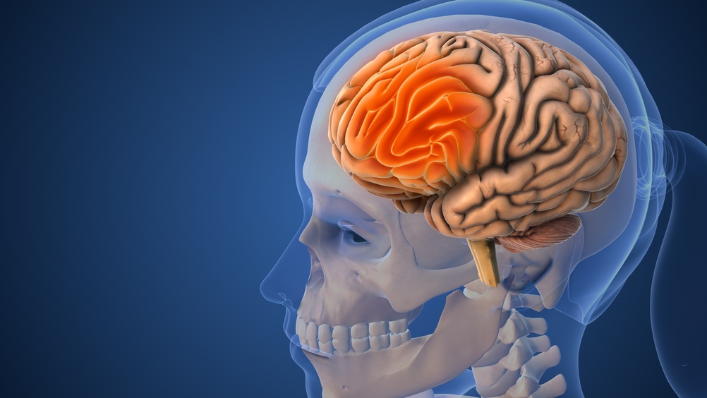
A structurally intact blood-brain barrier is a prerequisite for safe heavy metal chelation and aggressive detoxification. When that barrier is compromised, mobilized metals and toxins gain access to the central nervous system in concentrations that would otherwise be filtered out. The neurological consequences can be severe and rapid.
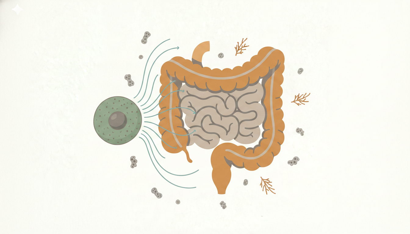
Clinical patterns suggesting blood-brain barrier permeability include a history of head trauma, chronic neuroinflammation, unresolved intestinal permeability with systemic immune activation, significant alcohol use, and chronic sleep deprivation. Laboratory markers such as elevated S100B or GFAP antibodies, when available and interpreted in clinical context, can support this assessment. The presence of neurological symptoms that worsen with chemical exposure is itself a clinical signal worth taking seriously.
Dr. Kharrazian's clinical teaching emphasizes that practitioners should evaluate blood-brain barrier status not as a secondary consideration but as a primary gating question before detox. If the barrier is compromised, the detox pathway changes entirely. The goal shifts to barrier support and restoration first — addressing intestinal permeability, reducing systemic inflammation, and improving sleep — before any mobilization protocol is considered.
Chemical Antibodies to Heavy Metals: What an Immune Response Is Telling You
The presence of chemical antibodies to heavy metals signals that the immune system has already mounted a specific response to those compounds. This matters clinically in a way that is frequently underappreciated.
An antibody response means the immune system has been sensitized. When chelation or detox mobilizes metals that have been stored in tissues, the sudden increase in circulating antigen can trigger a pronounced immune reaction — not the gradual clearance the practitioner intended, but an amplified response in a system that is already primed to react. Research in immunology identifies this as a form of antigen re-exposure in a sensitized host, and the outcomes can include significant inflammatory flares, autoimmune exacerbation, and worsening neurological symptoms.
Practically, testing for chemical antibodies before initiating detox gives the practitioner critical information. A patient with measurable antibody reactivity to mercury, lead, or other heavy metals is not a candidate for aggressive mobilization. The clinical strategy in this population requires immune modulation first — reducing systemic immune reactivity, supporting regulatory T-cell function, and addressing the barriers that allowed sensitization to develop — before any attempt to mobilize stored metals.
Primed Glial Cells and Why Neuroinflammatory Status Must Be Assessed Before Detox
Glial cells — microglia and astrocytes — are the immune cells of the central nervous system. Under normal conditions, they shift between resting and activated states as needed, then return to baseline. In a patient with chronic neuroinflammation, glial cells become primed: sensitized to a state of low-grade ongoing activation that generates disproportionate inflammatory responses to stimuli that would otherwise be inconsequential.
Primed glial cells do not require a large trigger to produce a large reaction. The oxidative stress generated by detoxification, or the neurological impact of mobilized toxins circulating in a compromised system, can be sufficient. The result is a worsening of the neuroinflammatory state the practitioner may have been trying to address in the first place.
Clinical indicators of primed glial cells include chronic brain fog, sensitivity to light or sound, poor alcohol tolerance, worsening cognitive function with minor physiological stressors (illness, poor sleep, dietary deviation), and a history of traumatic brain injury. These patients often describe feeling "wrecked" for days after what should be a minor physical or metabolic challenge. That disproportionate response is the clinical fingerprint of a primed neuroinflammatory state.
Dr. Kharrazian's coursework teaches practitioners to identify this pattern and prioritize neuroinflammation resolution before detox. This means addressing the inputs that sustain glial priming — blood sugar dysregulation, gut-brain immune axis dysfunction, ongoing infections, sleep disruption — and supporting glial regulatory mechanisms before any detox protocol begins.
The Clinical Decision: What to Do When a Patient Presents All Five Patterns
A patient who presents with physical frailty, cognitive decline, clinical signs of blood-brain barrier permeability, positive chemical antibody reactivity, and indicators of primed glial cells is not a candidate for detox. The question becomes: what is the appropriate clinical strategy?
The sequence of thought shifts from "how do we detox this patient" to "what needs to stabilize before detox becomes safe." That sequence typically involves reducing the total inflammatory burden, restoring barrier integrity at both the gut and the blood-brain barrier, supporting mitochondrial function and antioxidant capacity, modulating immune reactivity, and addressing the lifestyle and physiological inputs that are sustaining the fragility. For some patients, that work takes months. For others, systemic fragility is severe enough that aggressive mobilization protocols should never be on the table.
The practitioners who cause harm with detox are not reckless. They are often highly motivated clinicians pursuing the right goal through the wrong sequence. The assessment process Dr. Kharrazian's clinical training provides gives practitioners the thought process to know when to proceed, when to prepare the patient first, and when to pursue a different strategy entirely.
Key Takeaways
- Physical frailty in elderly patients reduces the metabolic and antioxidant reserve needed to process mobilized toxins safely.
- Dementia compromises both neurological tolerance and the patient's ability to report adverse reactions, making safe protocol management unreliable.
- A compromised blood-brain barrier converts detox mobilization from a clearance event into a potential neurological insult.
- Chemical antibody reactivity to heavy metals indicates immune sensitization — mobilizing those metals can trigger amplified inflammatory responses.
- Primed glial cells produce disproportionate neuroinflammatory responses to the oxidative and toxic load generated by detox protocols.
Frequently Asked Questions
A systemically fragile patient lacks the physiological reserve — antioxidant capacity, barrier integrity, immune regulation, and neurological stability — needed to safely process the demands detoxification creates. In these patients, mobilizing stored toxins can worsen the conditions the protocol was intended to address.
Clinical history is the primary tool: head trauma, chronic neuroinflammation, intestinal permeability, significant alcohol use, and chronic sleep deprivation all increase risk. Serum markers such as S100B and GFAP antibodies can support clinical assessment when available and interpreted in full clinical context.
Yes, but not before addressing immune sensitization. The clinical strategy requires reducing systemic immune reactivity and supporting regulatory immune function first. Attempting mobilization in a sensitized patient without this preparation significantly increases the risk of an exaggerated immune response.
Chronic brain fog, light and sound sensitivity, poor alcohol tolerance, disproportionate cognitive worsening after minor physiological stressors, and prolonged recovery from illness or injury. These indicate a neuroinflammatory state in which the central nervous system reacts excessively to inputs that healthy patients tolerate without consequence.
Stabilize first. Reduce total inflammatory burden, restore gut and blood-brain barrier integrity, support mitochondrial and antioxidant function, and modulate immune reactivity. Detoxification protocols are appropriate only after these foundational systems are functioning at a level sufficient to support safe mobilization and clearance.
About the Author
Dr. Datis Kharrazian, PhD, DHSc, DC, MS, MMSc, FACN, is a Harvard Medical School research fellow and researcher at Massachusetts General Hospital's Department of Neurology specializing in autoimmunity and neuroimmunology. He serves as Associate Clinical Professor at Loma Linda University School of Medicine and is the author of Why Do I Still Have Thyroid Symptoms When My Lab Tests Are Normal and Why Isn't My Brain Working. He is a Fellow of the American College of Nutrition, a Diplomate of the Board of Nutrition Specialists, a member of the American Association of Immunologists, and a Fellow of the Royal Society of Medicine (UK). The Kharrazian Institute provides clinical education and training to more than 5,000 physicians and healthcare providers worldwide.
The Kharrazian Institute's advanced clinical training covers detox readiness assessment, neuroinflammation protocols, and the clinical strategies needed to manage complex, chronic patients safely. Learn more about available courses at the Kharrazian Institute.