Most practitioners treating neurological cases are looking for the wrong thing. They're searching for the trigger they haven't identified yet — the infection, the toxin, the hormone imbalance — when the more clinically urgent problem is that the brain's immune system has already been structurally reorganized by previous insults. The trigger isn't the issue anymore. The microglia are.
Dr. Datis Kharrazian and the Kharrazian Institute address microglial activation clinical patterns extensively in advanced functional neurology training, because this is precisely where treatment-resistant cases stop responding to conventional approaches. Understanding what primed microglia actually do — and why they behave differently than newly activated ones — changes how practitioners sequence their clinical thinking.
What Microglial Activation Actually Means Clinically
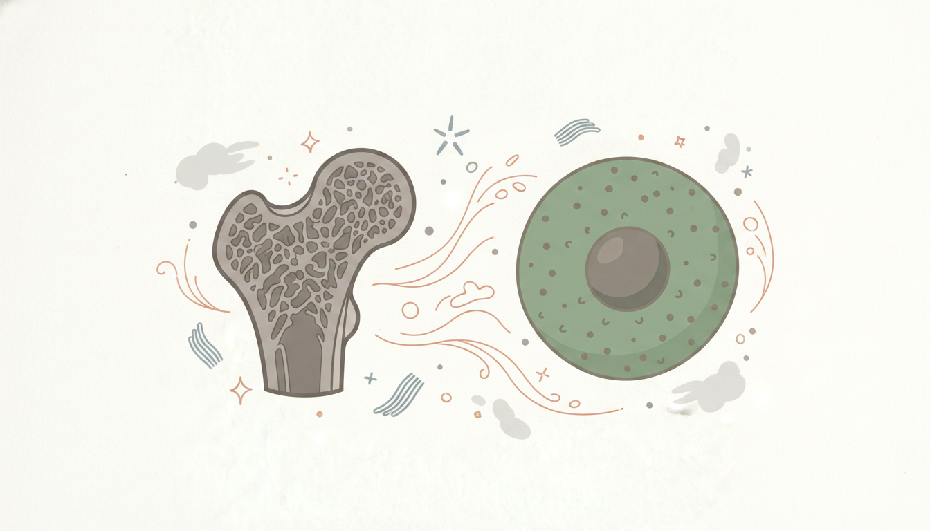
Microglia are the resident immune cells of the central nervous system. Under normal conditions, they perform surveillance, pruning synapses, clearing debris, and responding to injury with a controlled inflammatory response. The problem is that "controlled" depends entirely on the state of the microglia before a new trigger arrives.
Research in neuroimmunology identifies two functional activation states. M1 activation is pro-inflammatory: microglia release cytokines including TNF-α, IL-1β, and IL-6, creating the neuroinflammatory environment responsible for symptoms like brain fog, cognitive slowing, heightened pain sensitivity, mood dysregulation, and fatigue. M2 activation is anti-inflammatory and reparative: microglia release IL-10 and TGF-β and support tissue remodeling. Neither state is inherently pathological. The problem is when M1 activation becomes chronic, disproportionate, or self-sustaining.
Dr. Kharrazian's clinical training frames this as a signal-to-noise problem. A brain with healthy microglial function responds to a legitimate stressor, mounts an M1 response, resolves it, and returns to surveillance mode. A brain with primed microglia responds to nearly any input — physiological stress, a minor infection, blood sugar dysregulation, a disrupted sleep cycle — as though it were a major threat. The amplitude of the inflammatory response no longer correlates with the severity of the trigger.
Glial Priming: Why Past Injuries Don't Stay in the Past
Glial priming is the mechanism that explains why so many neurological cases become treatment-resistant over time. A single significant neuroinflammatory event — traumatic brain injury, CNS infection, severe metabolic crisis, or prolonged systemic inflammation — can permanently alter microglial sensitivity thresholds. The cells remain in a low-grade activated state. They don't return to baseline.
When a primed microglial population encounters a new trigger, the inflammatory response is exaggerated and prolonged. Research in neuroinflammation describes this as a "second-hit" phenomenon: the first insult doesn't cause overt pathology, but it lowers the threshold so dramatically that a second, otherwise subclinical, insult produces a disproportionate neurological response. This is why patients with a history of concussion, childhood meningitis, or Lyme disease often present with neurological symptoms that seem far out of proportion to their current clinical picture.
Dr. Kharrazian's coursework teaches practitioners to treat the patient's history as the primary diagnostic document in these cases. A thorough timeline of neurological insults — including events the patient may not consider medically significant, like multiple sports concussions or a severe case of COVID-19 years prior — often reveals the priming event that makes the current presentation legible. Without that history, practitioners end up chasing the most recent trigger while the underlying sensitization goes unaddressed.
Identifying Microglial Activation Clinical Patterns in Practice
Primed microglia produce a recognizable symptom signature, though it's rarely labeled correctly in conventional workups. Patients with active microglial-driven neuroinflammation typically present with central sensitization — widespread pain with no structural explanation, thermal dysregulation, heightened sensitivity to light and sound, and cognitive symptoms that fluctuate with physical or emotional stressors. The variability is clinically confusing unless the practitioner knows what to look for.
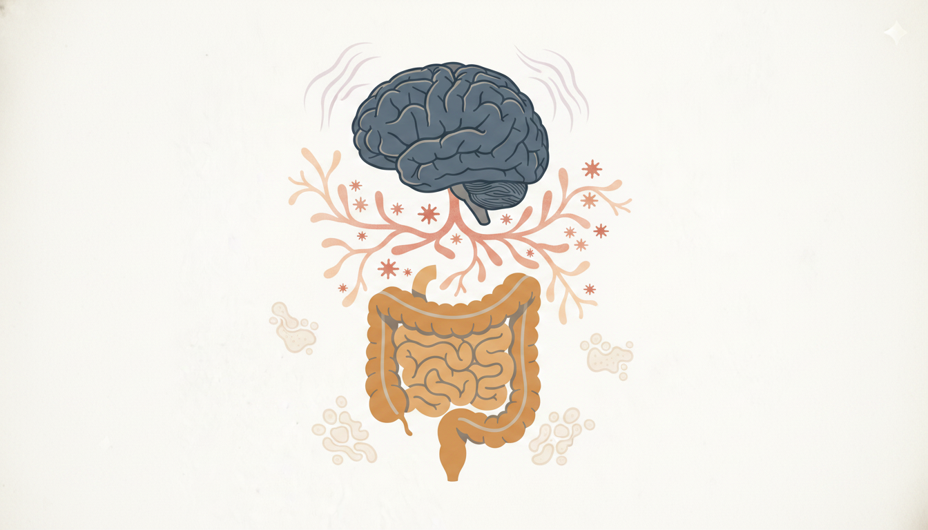
Several triggers consistently activate primed microglia. Hypoxia is among the most clinically underappreciated: poor sleep, sleep apnea, and cardiovascular compromise all reduce cerebral oxygen delivery and can sustain M1 activation independent of any immune trigger. Systemic infections reliably cross-activate CNS immunity through cytokine signaling. Blood-brain barrier disruption, whether from intestinal permeability, chronic stress, or direct CNS injury, allows peripheral inflammatory signals to reach microglial cells directly. Blood glucose dysregulation produces oxidative stress in neural tissue. Each of these can serve as a recurring activation trigger in a patient whose microglia are already primed.
The clinical pattern that signals microglial priming rather than straightforward neuroinflammation is disproportionate reactivity. The patient who crashes neurologically after a minor illness, whose cognitive symptoms flare with emotional stress, or who has never fully recovered baseline function after a distant neurological event — that patient's clinical trajectory points toward priming, not acute activation.
Why Treatment-Resistant Neuroinflammation Requires a Different Clinical Strategy
Standard anti-inflammatory protocols frequently fail in primed cases because they address the downstream output of M1 activation without addressing the sensitized state itself. Reducing systemic inflammation will not normalize a microglial population that responds disproportionately to every input. The clinical strategy has to address the sensitization threshold directly.
Dr. Kharrazian's advanced functional neurology training outlines a sequenced approach for these cases. The first priority is identifying and eliminating the recurring triggers that continue to drive activation. Hypoxia sources, blood glucose instability, blood-brain barrier disruption, and ongoing systemic infections each need to be assessed and addressed systematically. If a patient's microglia are being continuously reactivated by nocturnal hypoxia, for example, no neuroprotective protocol will produce durable results.
The second priority is supporting the shift from M1 to M2 activation states. Research in neuroinflammation has identified several compounds with documented ability to cross the blood-brain barrier and modulate microglial phenotype. Flavonoids — including luteolin and apigenin — reduce pro-inflammatory cytokine output from activated microglia. Magnesium supports neuronal function and reduces excitotoxicity, which independently sustains microglial activation. Omega-3 fatty acids, specifically DHA, are incorporated into neural membranes and shift microglial signaling toward resolution. The clinical application of these compounds requires both appropriate dosing and timing relative to the activation state — something Dr. Kharrazian's coursework addresses in protocol-level detail.
Neuromodulation strategies occupy the third tier. Aerobic exercise promotes neuroplasticity and has documented effects on microglial M2 polarization through BDNF upregulation and anti-inflammatory myokine release. Activation of descending inhibitory pain pathways reduces central sensitization, which otherwise sustains the neuroinflammatory signal. These strategies are not adjuncts in treatment-resistant cases — they are often the primary mechanism through which the sensitization threshold can be raised.
Long-Term Management When Priming Has Occurred
Permanent microglial priming changes the therapeutic goal. The aim is no longer resolution — it's management of the threshold. Primed patients require ongoing monitoring because their neurological stability depends on continuous avoidance of trigger accumulation. A single significant stressor can reset months of clinical progress.
This has direct implications for how practitioners communicate with these patients. The explanation that "your brain's immune cells have a lower threshold after past injuries" is both accurate and practically actionable. It reframes why lifestyle consistency — sleep quality, stress load, glycemic control, physical activity — functions as an ongoing neuroprotective strategy rather than a general wellness recommendation. Patients who understand the mechanism tend to maintain the behaviors that support it.
The research synthesized in Dr. Kharrazian's functional neurology coursework also addresses the monitoring side: tracking symptom variability relative to known triggers, using neurological function assessments to detect early regression, and adjusting protocols before full decompensation occurs rather than after. In primed cases, reactive management is consistently less effective than the anticipatory kind.
The Clinical Opportunity in Treatment-Resistant Cases
Treatment-resistant neuroinflammation is not an absence of clinical options. It's a presentation that requires a more precise clinical strategy than most practitioners have been trained to apply. The research base in neuroimmunology supporting microglial priming as a mechanism is substantial — the clinical gap is in translating that research into actionable protocols for the patients sitting in an office today.
Dr. Kharrazian's advanced functional neurology training exists specifically to close that gap. The practitioners seeing the best outcomes in complex neurological cases are the ones who understand not just that neuroinflammation is present, but which activation pattern they're dealing with, how long it has been operating, and what the brain's actual sensitization state is. That distinction drives every clinical decision that follows.
Key Takeaways
- Glial priming permanently lowers microglial activation thresholds following significant neuroinflammatory events, producing disproportionate responses to subsequent triggers.
- Distinguishing between acute M1 activation and a primed microglial state is essential before selecting a treatment protocol, because the two require different clinical strategies.
- Hypoxia, blood glucose dysregulation, blood-brain barrier disruption, and systemic infections are the most clinically common recurring triggers sustaining neuroinflammation in primed patients.
- Blood-brain barrier-crossing nutraceuticals including flavonoids, magnesium, and DHA support M2 polarization and can be used adjunctively to reduce M1 output.
- In primed cases, the therapeutic goal shifts from resolution to threshold management — requiring ongoing monitoring and consistent trigger reduction rather than episodic intervention.
Frequently Asked Questions
Glial priming occurs when prior neuroinflammatory events permanently sensitize microglial cells, lowering their activation threshold. Primed microglia mount exaggerated inflammatory responses to minor triggers, which is why these patients show neurological symptoms disproportionate to current stressors and fail to respond to standard anti-inflammatory protocols.
M1 activation is pro-inflammatory and produces cytokines associated with brain fog, pain sensitization, cognitive slowing, and mood changes. M2 activation is anti-inflammatory and supports tissue repair. Chronic M1 dominance, particularly in primed microglia, sustains neurological symptoms independent of the original trigger and requires targeted clinical intervention to shift.
The most clinically significant recurring triggers are hypoxia from poor sleep or sleep apnea, blood glucose dysregulation causing oxidative neural stress, blood-brain barrier disruption from intestinal permeability or direct CNS injury, and systemic infections activating central immunity through peripheral cytokine signaling.
Current neuroimmunology research suggests structural changes from glial priming are largely permanent, though the degree of activation and symptom severity can be significantly reduced. The clinical goal becomes threshold management — identifying and minimizing recurring triggers and supporting M2 polarization — rather than expecting complete resolution of the sensitized state.
Research in neuroinflammation identifies flavonoids such as luteolin and apigenin, DHA from omega-3 sources, and magnesium as compounds with documented blood-brain barrier penetration and microglial modulatory effects. Clinical application requires appropriate dosing and protocol sequencing based on the patient's current activation state.
About the Author
Dr. Datis Kharrazian, PhD, DHSc, DC, MS, MMSc, FACN is a Harvard Medical School research fellow and researcher at Massachusetts General Hospital's Department of Neurology, specializing in autoimmunity and neuroimmunology. He serves as Associate Clinical Professor at Loma Linda University School of Medicine and is the author of Why Do I Still Have Thyroid Symptoms When My Lab Tests Are Normal and Why Isn't My Brain Working. He is a Fellow of the American College of Nutrition, a Diplomate of the Board of Nutrition Specialists, a member of the American Association of Immunologists, and a Fellow of the Royal Society of Medicine (UK). The Kharrazian Institute serves more than 5,000 physicians and healthcare providers worldwide.
Dr. Kharrazian's advanced functional neurology training covers microglial activation clinical patterns, glial priming mechanisms, and protocol-level strategies for treatment-resistant neuroinflammation. Practitioners managing complex neurological cases can access this training through the Kharrazian Institute.
Explore Functional Neurology Courses at the Kharrazian Institute