Most practitioners treating chronic pain are looking at the wrong organ. The joints, the fascia, the gut — these are common starting points. But when a patient's pain doesn't resolve despite thorough musculoskeletal and inflammatory workups, the brain itself may be the source of dysregulation. This is a clinical reality that Dr. Datis Kharrazian and the Kharrazian Institute address directly in their advanced neurology training for practitioners: traumatic brain injury pain isn't just pain that originates in the brain. It's pain the brain has lost the ability to suppress.
That distinction changes everything about how you assess and manage these patients.
How Brain Injury Disrupts Pain Modulation
The brain doesn't just receive pain signals. It actively dampens them. Descending inhibitory pathways — running from cortical and subcortical structures down through the brainstem and spinal cord — continuously regulate how much nociceptive input reaches conscious awareness. A healthy brain filters. An injured brain filters poorly.
After traumatic brain injury, those inhibitory pathways are often compromised. The result is central sensitization: a state where the central nervous system amplifies pain signals disproportionate to the actual tissue input. The patient reports severe pain. Imaging finds nothing structural. Labs are unremarkable. The practitioner concludes the problem is psychological. The real problem is neurological.
Research in neuroimmunology demonstrates that this sensitization isn't just functional — it has a clear biochemical substrate. Injured neural tissue triggers microglial activation, and activated microglia release pro-inflammatory cytokines that lower the threshold for nociceptor activation throughout the central nervous system. Pain becomes easier to generate and harder to turn off. This is the mechanism Dr. Kharrazian's clinical coursework teaches practitioners to identify when the standard workup comes back clean but the patient cannot recover.
The Role of Neuroinflammation in Chronic Pain After TBI
Neuroinflammation and chronic pain are not parallel problems after a brain injury. One drives the other.
Microglial cells — the brain's resident immune cells — enter a primed state following trauma. In a primed state, they don't return to baseline after the initial injury resolves. They remain hyperreactive, releasing inflammatory mediators in response to stimuli that would not have triggered a response in uninjured tissue. Every subsequent stressor — infection, metabolic insult, sleep deprivation, psychological stress — amplifies the neuroinflammatory environment and, with it, pain sensitivity.
Research in neuroinflammation and chronic pain shows that this glial priming can persist for years, sometimes decades, after the original injury. A patient presenting with diffuse pain syndromes, cognitive fog, and fatigue a decade after a concussion is not an anomaly. They are showing the predictable downstream effects of unresolved neuroinflammation. Dr. Kharrazian's coursework at KI trains practitioners to connect this timeline — to ask not just what is happening now, but what happened to the brain years or decades prior.
Sympathetic Dysregulation and the Pain-Stress Loop
Brain injuries disrupt more than pain pathways. They impair the brain's regulation of the autonomic nervous system, and that dysregulation feeds directly back into pain.
The prefrontal cortex and limbic structures exert significant control over sympathetic tone. When those areas are damaged or chronically inflamed, sympathetic output becomes dysregulated — often elevated. Elevated sympathetic tone drives increased catecholamine release. Catecholamines sensitize peripheral nociceptors. The patient feels more pain, the pain increases stress, and the stress amplifies sympathetic output further. The loop is self-sustaining without intervention at the neurological level.
This is why standard pain management approaches often fail these patients. Anti-inflammatories address peripheral inflammation. They don't address centrally driven sympathetic dysregulation. Addressing traumatic brain injury pain requires working upstream — at the level of autonomic regulation and brain function — not just at the site where the patient reports discomfort.
Neurovascular Coupling and Why It Matters for Pain
Neurovascular coupling is the brain's ability to match blood flow to metabolic demand in active neural regions. When a region fires, blood flow should increase to meet the energy requirement. After TBI, this coupling is frequently impaired.
Impaired neurovascular coupling creates regional energy deficits in brain tissue. Neurons in energy-deficient states cannot sustain normal inhibitory output. Inhibitory interneurons — the cells responsible for dampening excitatory signals — are particularly vulnerable to ATP deficits because inhibition is metabolically expensive. When those interneurons fail, excitatory pathways become disinhibited. Pain pathways stay open. The patient experiences chronic, diffuse pain that fluctuates with metabolic state.
Neurovascular coupling assessment gives practitioners a window into this mechanism. Dr. Kharrazian's advanced neurology training covers the clinical tools for evaluating neurovascular coupling and interpreting what impaired coupling means for the patient's symptom picture — including pain that seems to have no peripheral origin.
Mitochondrial Dysfunction as a Pain Amplifier
Brain injury consistently impairs mitochondrial function in neural tissue. The bioenergetic consequences extend well beyond fatigue.
ATP is required for nearly every aspect of normal neurological function: maintaining ion gradients, sustaining membrane potential, driving inhibitory neurotransmission. When mitochondrial output drops, neurons struggle to maintain these functions. The threshold for nociceptor activation drops with them. Signals that wouldn't register as painful under normal bioenergetic conditions now breach threshold. This is the mechanism by which mitochondrial dysfunction amplifies pain perception — not through inflammation alone, but through direct energetic failure of the inhibitory machinery.
Research in cellular neuroscience shows that injured mitochondria also generate excess reactive oxygen species, which further sensitize pain pathways and sustain neuroinflammation. Mitochondrial dysfunction and neuroinflammation are not independent variables in post-TBI pain — they are mutually reinforcing. A clinical strategy that addresses one while ignoring the other will produce incomplete results.
The Brain-Gut Axis: A Bidirectional Complication
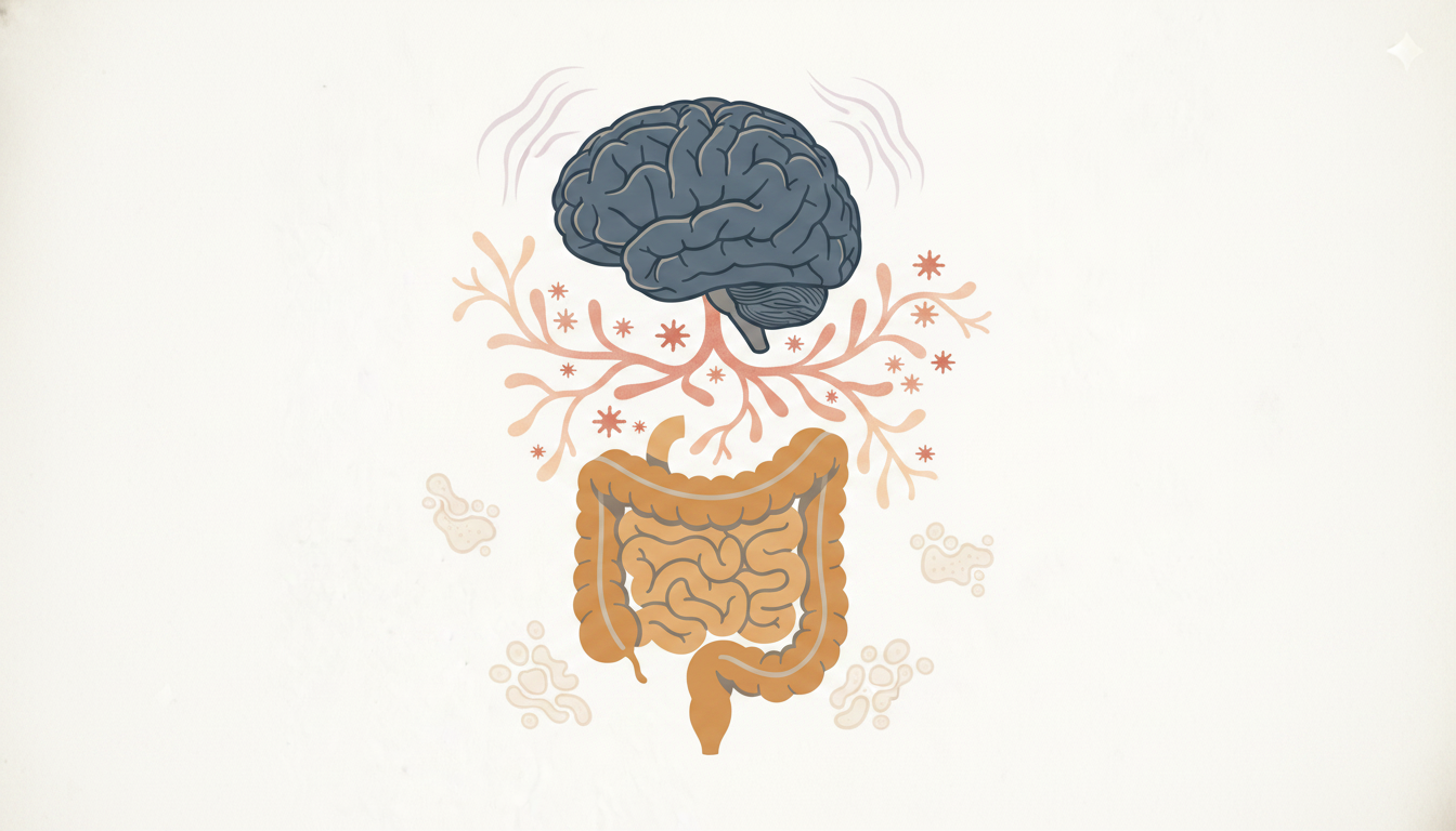
Chronic neuroinflammation after TBI doesn't stay in the brain. Through the brain-gut axis, it alters gut motility, intestinal permeability, and the composition of the gut microbiome. Gut dysbiosis and intestinal permeability then generate systemic inflammatory signals that travel back up the vagus nerve and amplify neuroinflammation centrally.
This bidirectional loop means that a patient with unresolved TBI symptoms may be sustaining their own neuroinflammation through gut dysfunction that developed as a consequence of the original brain injury. Brain-gut axis dysfunction in post-TBI patients is not a coincidence. It is a predictable consequence of chronic autonomic dysregulation and neuroinflammation.
Practitioners who treat the gut without addressing the brain, or the brain without addressing the gut, are working with an incomplete picture. Dr. Kharrazian's clinical training addresses this bidirectional relationship and teaches practitioners how to sequence interventions when both systems are compromised simultaneously.
What Clinical Management Actually Requires
Managing traumatic brain injury pain requires addressing the underlying mechanisms driving central sensitization — not suppressing the pain signal while those mechanisms continue unchecked.
Optimizing brain bioenergetics is foundational. Without adequate ATP production, the inhibitory pathways that modulate pain cannot function, regardless of what anti-inflammatory strategies are in place. Mitochondrial support, neurovascular coupling, and cerebral blood flow are legitimate clinical targets for this population.
Resolving neuroinflammation is equally non-negotiable. Research in neuroinflammation and chronic pain consistently shows that unresolved glial activation perpetuates central sensitization. Identifying and removing the triggers maintaining microglial priming — whether metabolic, dietary, infectious, or environmental — is a core component of any protocol designed to produce durable improvement.
Finally, autonomic regulation must be assessed. If sympathetic dysregulation is sustaining peripheral nociceptor sensitization, addressing only central inflammation will leave a significant driver of pain untreated. The clinical strategy has to account for the full loop: brain injury, neuroinflammation, autonomic dysregulation, peripheral sensitization, and the gut-brain feedback that sustains the cycle.
Key Takeaways
- Traumatic brain injury impairs the brain's descending inhibitory pathways, producing central sensitization and chronic pain disproportionate to peripheral tissue findings.
- Microglial priming after TBI sustains neuroinflammation long after the original injury, continuously lowering the threshold for pain signal generation.
- Sympathetic dysregulation from brain injury creates a self-sustaining pain-stress loop that standard pain management approaches do not address.
- Mitochondrial dysfunction reduces ATP availability, directly impairing the inhibitory neurotransmission that keeps pain pathways from becoming chronically active.
- Brain-gut axis dysfunction in post-TBI patients is a predictable consequence of neuroinflammation and autonomic dysregulation, not a coincidental comorbidity.
Frequently Asked Questions
Microglial priming after TBI keeps neuroinflammatory pathways active well beyond the initial injury window. This sustained inflammation continuously sensitizes pain pathways and impairs the brain's descending inhibitory control, producing chronic pain that persists without an obvious ongoing peripheral cause.
Neuroinflammation lowers the threshold for nociceptor activation by releasing pro-inflammatory cytokines that sensitize pain pathways throughout the central nervous system. The result is central sensitization — amplified pain perception that cannot be resolved by treating only the peripheral site where pain is reported.
Inhibitory neurotransmission is metabolically expensive. When mitochondrial dysfunction reduces ATP production in neural tissue, inhibitory interneurons fail first. Pain pathways become disinhibited, and the threshold for nociceptor activation drops — meaning stimuli that would not normally produce pain now breach the activation threshold.
Neurovascular coupling is the brain's ability to increase blood flow to active neural regions on demand. When this is impaired after TBI, active brain regions develop energy deficits that compromise normal inhibitory function. In pain management, this means brain regions responsible for dampening pain signals cannot sustain that output.
Yes. Chronic neuroinflammation and autonomic dysregulation following TBI impair gut motility, increase intestinal permeability, and alter microbiome composition. The resulting gut dysfunction then generates systemic inflammatory signals that travel back to the brain via the vagus nerve, sustaining the neuroinflammation driving the original symptoms.
About the Author
Dr. Datis Kharrazian, PhD, DHSc, DC, MS, MMSc, FACN, is a Harvard Medical School research fellow and researcher at Massachusetts General Hospital's Department of Neurology specializing in autoimmunity and neuroimmunology. He serves as Associate Clinical Professor at Loma Linda University School of Medicine and is the author of Why Do I Still Have Thyroid Symptoms When My Lab Tests Are Normal and Why Isn't My Brain Working. He is a Fellow of the American College of Nutrition, Diplomate of the Board of Nutrition Specialists, member of the American Association of Immunologists, and Fellow of the Royal Society of Medicine (UK). The Kharrazian Institute provides clinical education and training to more than 5,000 physicians and healthcare providers worldwide.
Advance Your Clinical Approach to TBI and Chronic Pain
The Kharrazian Institute's neurology training covers the assessment and management of central sensitization, neuroinflammation, neurovascular coupling, and autonomic dysregulation in patients with complex chronic conditions. Practitioners seeking evidence-based clinical strategies for TBI-related chronic pain syndromes can explore current course offerings at the Kharrazian Institute.