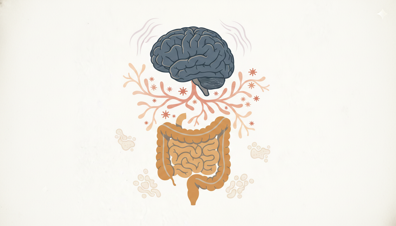
Most practitioners treating hypertension are not asking about gut symptoms. Most practitioners managing post-TBI patients are not running cardiovascular risk assessments. That clinical separation is the problem. The brain, gut, and vascular system are not independent targets — they are linked through neuroimmunological and neuroendocrine pathways that, when disrupted, create a cascade of dysfunction across all three simultaneously. Dr. Datis Kharrazian addresses this intersection directly in his clinical training at the Kharrazian Institute, where the brain-gut vascular dysfunction connection is taught as an integrated clinical strategy rather than a collection of separate organ problems.
How Brain Injury Disrupts Gut Function Through the Vagal Pathway
Traumatic brain injury (TBI) and cerebrovascular events do not stay in the brain. Within hours of a significant brain injury, measurable changes in intestinal permeability occur. This is not a secondary complication — it is an immediate physiological consequence of how brain injury disrupts autonomic signaling to the gut.
The gut-brain axis depends on intact neuroendocrine and neuroimmunological communication through regions including the hypothalamus, insular cortex, and anterior cingulate cortex. These areas regulate gastrointestinal motility, secretory function, and mucosal barrier integrity via vagal efferent pathways. When TBI disrupts these regions, vagal output to the gut drops. The enteric nervous system loses its upstream regulatory signal, and the gut responds predictably: motility slows, mucosal barrier function weakens, and intestinal permeability increases.
The clinical implication is direct. A patient with a history of TBI who presents with chronic bloating, altered motility, or unexplained gastrointestinal symptoms may not have a primary gut condition. The gut is responding to a loss of central regulation. Treating the gut alone will produce incomplete results.
Intestinal Permeability and the Vascular Inflammatory Cascade
Once intestinal permeability increases following brain injury, the barrier between luminal contents and systemic circulation is compromised. Lipopolysaccharides (LPS) — gram-negative bacterial cell wall components — translocate into circulation. This is the mechanism connecting the gut to the vascular system in ways most practitioners never assess.
Circulating LPS activates toll-like receptor 4 (TLR4) on endothelial cells, triggering nuclear factor kappa B (NF-κB) signaling and the release of pro-inflammatory cytokines including TNF-α, IL-1β, and IL-6. These cytokines directly damage vascular endothelium, increase arterial stiffness, and impair nitric oxide-mediated vasodilation. Research in vascular immunology demonstrates that chronic low-level endotoxemia is an independent risk factor for atherosclerosis and cardiovascular events — a connection Dr. Kharrazian's coursework teaches practitioners to identify in patients whose cardiovascular risk is disproportionate to their conventional risk factors.
The gut, in this model, is not a bystander. It is an active source of vascular inflammatory burden when the brain can no longer regulate its barrier function.
Autonomic Dysregulation, Sympathetic Overactivity, and Hypertension
Brain injury that affects autonomic regulatory centers produces dysautonomia. In practical terms, that means sympathetic overactivity becomes the default state. Sympathetic dominance raises resting heart rate, increases peripheral vascular resistance, and elevates blood pressure through catecholamine-mediated mechanisms.
Patients with post-TBI autonomic dysfunction frequently develop hypertension that does not respond well to standard antihypertensive strategies. The elevated blood pressure is not a primary vascular disease — it is a downstream consequence of dysregulated central autonomic output. Practitioners who treat the blood pressure without addressing the underlying autonomic imbalance are managing a symptom, not the cause.
Sympathetic overactivity simultaneously worsens gut function. Elevated sympathetic tone suppresses vagal activity further, reducing gut motility, reducing secretory IgA output, and impairing mucosal repair. The autonomic dysfunction creates a self-reinforcing loop: brain injury disrupts autonomic regulation, sympathetic overactivity suppresses vagal gut regulation, gut barrier integrity degrades further, LPS load increases, vascular inflammation rises, and cerebrovascular perfusion is increasingly compromised. Each element worsens the others.
Substance P, Neurogenic Inflammation, and Vascular Damage
TBI triggers the release of substance P from peripheral and central neurons. Substance P is a neuropeptide with direct vascular effects: it increases vascular permeability, promotes plasma extravasation, and activates mast cells. In the context of a compromised gut barrier, substance P release compounds mucosal inflammation and amplifies the systemic inflammatory signal reaching the vascular endothelium.
Research in neuroimmunology identifies substance P as a key mediator connecting neurological injury to systemic vascular inflammation. Elevated substance P following TBI is associated with increased blood-brain barrier permeability, worsened neuroinflammation, and heightened cardiovascular risk. The clinical picture this creates — a patient with brain injury, gut complaints, and cardiovascular complications — is not a coincidence of three separate conditions. It is a single pathophysiological cascade with three visible endpoints.
What the Gut-Vascular Connection Looks Like in Practice
The practitioner sees a patient with a history of TBI from three years prior. The patient has persistent bloating, unpredictable bowel function, newly elevated blood pressure, and fatigue that does not improve with rest. Previous workups for each complaint have returned inconclusive results. Each specialist has ruled out disease in their own system and discharged the patient.
None of the individual workups are wrong. What is missing is the connecting mechanism. The clinical training Dr. Kharrazian provides teaches practitioners to read these symptoms as a coherent pattern: compromised vagal regulation driving intestinal permeability, LPS-mediated endothelial inflammation driving vascular dysfunction, and autonomic imbalance sustaining the entire cycle.
Practical clinical strategies in this population include assessing vagal tone through heart rate variability, evaluating markers of intestinal permeability and endotoxin load, supporting mucosal barrier integrity to reduce systemic LPS translocation, and addressing autonomic dysregulation as a primary target rather than an incidental finding. The sequence matters. Targeting vascular inflammation without reducing the gut-sourced inflammatory input produces incomplete and often temporary improvement.
Why Treating These Systems Separately Fails the Patient
The conventional model assigns organ ownership to specialists. Neurology manages the brain injury. Gastroenterology manages the gut complaints. Cardiology manages the blood pressure. Each assessment is thorough within its boundary. But the pathology does not respect those boundaries, and the treatments applied within each silo may actively work against each other.
Antihypertensives that reduce cardiac output may further impair cerebrovascular perfusion in a post-TBI brain already struggling with circulation. Prokinetic agents for gut motility do not address the central reason motility is disrupted. Anti-inflammatory strategies applied to the gut without accounting for the ongoing autonomic dysregulation remove one input into the inflammatory cascade while leaving others intact.
Research in both neuroimmunology and cerebrovascular gut health demonstrates that integrated assessment of the brain-gut-vascular axis produces meaningfully different clinical outcomes in chronic patients who cannot recover under fragmented care. Dr. Kharrazian's clinical training builds that integrative sequence of thought — not as an abstract concept, but as an immediately applicable clinical strategy for the complex cases practitioners see every week.
Key Takeaways
- TBI disrupts vagal output to the gut through damage to the hypothalamus, insular cortex, and cingulate cortex, causing immediate increases in intestinal permeability.
- LPS translocation from a compromised gut barrier activates vascular endothelial inflammation through TLR4 signaling, creating a direct gut-to-vascular inflammatory pathway.
- Post-TBI sympathetic overactivity suppresses vagal regulation further, worsening both gut barrier function and blood pressure control simultaneously.
- Substance P released after brain injury increases vascular permeability and amplifies systemic inflammation, connecting neurological injury to cardiovascular risk.
- Effective clinical management addresses the autonomic and intestinal permeability mechanisms first — not the downstream vascular symptoms alone.
Frequently Asked Questions
Yes. TBI disrupts vagal pathways that regulate gut motility, secretion, and mucosal integrity. Research in neuroimmunology shows that intestinal permeability increases within hours of significant brain injury, contributing to systemic inflammation and chronic gastrointestinal dysfunction in post-TBI patients.
Increased intestinal permeability allows LPS to enter circulation, activating TLR4 receptors on vascular endothelium and triggering inflammatory cytokines that impair vasodilation and increase arterial stiffness. Chronic low-level endotoxemia is an independent contributor to elevated blood pressure and cardiovascular risk.
Dysautonomia refers to dysregulation of the autonomic nervous system, commonly characterized by sympathetic overactivity. Elevated sympathetic tone reduces vagal output to the gut, slowing motility, impairing mucosal repair, and reducing secretory IgA — all of which worsen intestinal permeability and systemic inflammatory load.
Multiple mechanisms converge after TBI: autonomic dysregulation raises sympathetic tone and blood pressure, substance P release increases vascular permeability, and gut-derived LPS drives vascular endothelial inflammation. Together these create sustained cardiovascular risk that originates neurologically, not primarily in the vascular system.
Reducing intestinal permeability decreases the systemic LPS burden that drives endothelial inflammation. Research demonstrates that lowering circulating endotoxin levels reduces TLR4 activation, decreases pro-inflammatory cytokine output, and supports improved vascular endothelial function — addressing one major source of vascular inflammatory input.
About the Author
Dr. Datis Kharrazian, PhD, DHSc, DC, MS, MMSc, FACN is a Harvard Medical School research fellow and researcher at Massachusetts General Hospital Department of Neurology specializing in autoimmunity and neuroimmunology. He serves as Associate Clinical Professor at Loma Linda University School of Medicine and is the author of Why Do I Still Have Thyroid Symptoms When My Lab Tests Are Normal and Why Isn't My Brain Working. He is a Fellow of the American College of Nutrition, Diplomate of the Board of Nutrition Specialists, member of the American Association of Immunologists, and Fellow of the RoyalSociety of Medicine (UK). The Kharrazian Institute serves more than 5,000 physicians and healthcare providers worldwide.
Learn more about how Dr. Kharrazian's clinical training addresses brain-gut-vascular dysfunction in complex and chronic patient cases. Visit the Kharrazian Institute to explore available courses and practitioner programs.