A common practitioner mistake: removing gluten from an autoimmune patient's diet, watching them improve initially, then losing the clinical thread when symptoms return weeks later. The assumption is compliance failure. The more likely explanation is cross-reactivity — the patient's immune system has been trained to attack a molecular pattern, and that pattern exists in more than one food.
Autoimmune cross-reactivity is one of the more underappreciated drivers of treatment resistance in chronic autoimmune cases. At the Kharrazian Institute, Dr. Datis Kharrazian's clinical training teaches practitioners how to identify cross-reactivity patterns systematically, so dietary and environmental interventions actually hold.
What Molecular Mimicry Actually Does to the Immune System
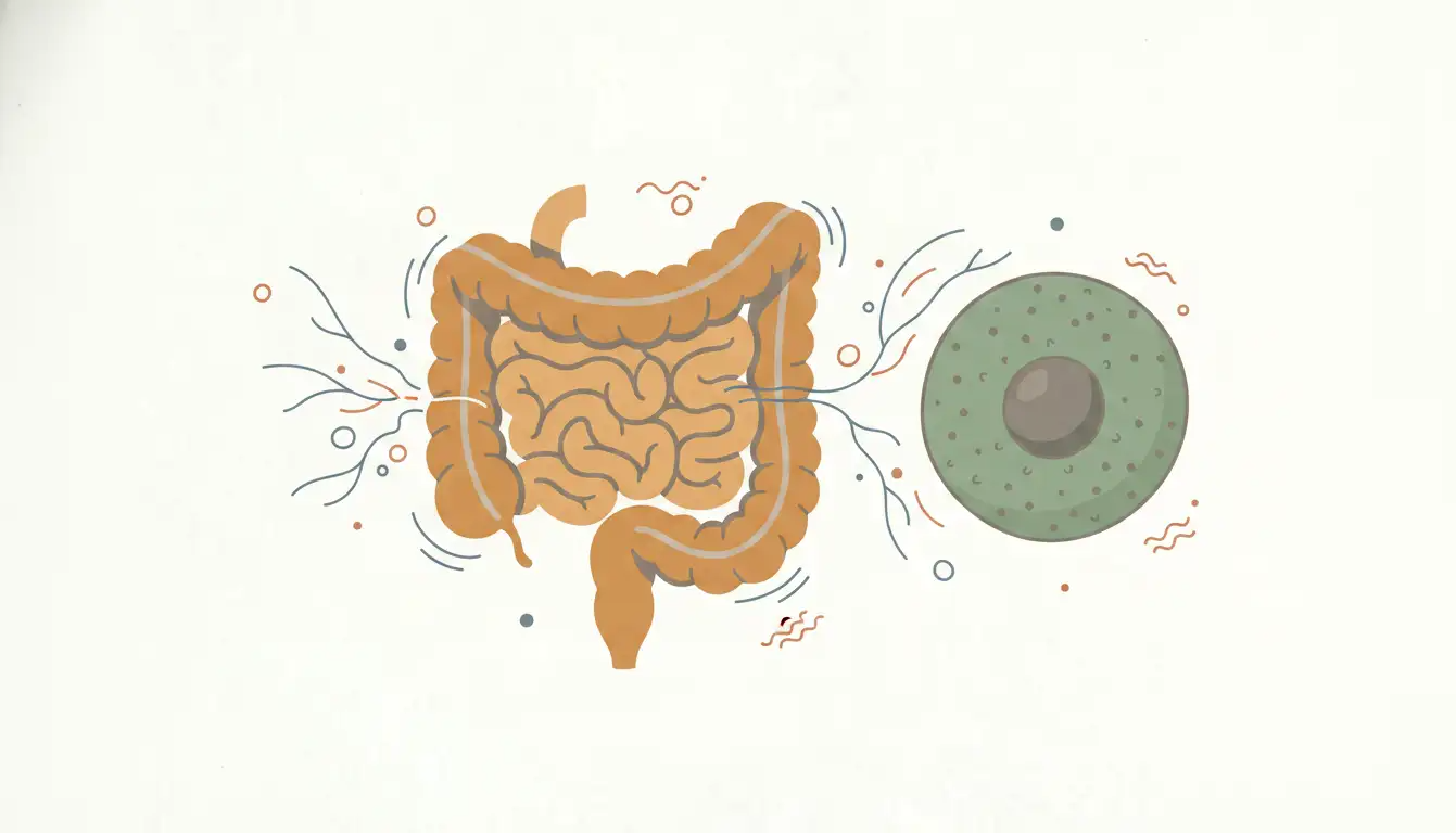
Molecular mimicry is the mechanism by which a foreign antigen — a protein from food, bacteria, or an environmental compound — shares enough structural similarity with a host tissue antigen that the immune system's antibodies attack both. The immune system isn't malfunctioning in a random way. It learned a pattern, and it's applying that pattern consistently.
Research in autoimmune immunology has established that gliadin, the immunogenic protein in gluten, contains peptide sequences structurally similar to several human tissue antigens. In genetically susceptible individuals, gliadin exposure doesn't just trigger local intestinal inflammation. The antibodies generated can cross-react with cerebellar tissue, thyroid tissue, and other targets depending on the individual's genetic antigen presentation. This is why two patients with the same gluten sensitivity can present with entirely different autoimmune conditions.
The clinical implication is direct: the trigger and the tissue damage don't have to be in the same anatomical location. A gut-level antigen exposure can drive neurological autoimmunity. Practitioners who treat only the local symptom picture will keep chasing a moving target.
Why Gluten Is Not the Only Dietary Cross-Reactor
Gluten receives the most clinical attention, but research in food immunology has identified a broader set of dietary proteins with cross-reactive potential. Dairy proteins, particularly casein and whey fractions, share structural homology with gliadin peptides. This is why a subset of gluten-sensitive patients see only partial improvement on a gluten-free diet — they've removed one antigen but left another with overlapping epitopes in the diet.
Other identified cross-reactive foods include corn, oats (even certified gluten-free varieties in sensitive individuals), yeast, and certain legumes. The cross-reactivity isn't universal across all patients — it depends on which antibodies the individual has generated and which tissue antigens those antibodies are targeting. This is where population-level dietary advice breaks down and personalized clinical assessment becomes necessary.
Dr. Kharrazian's clinical training emphasizes that practitioners shouldn't operate from a fixed elimination list. The relevant question for any given patient is: which antigens has this immune system learned to attack, and where else does that molecular pattern appear? That sequence of thought changes the clinical approach entirely.
How Cross-Reactive Antibodies Drive Ongoing Tissue Damage
Once the immune system has generated antibodies against a self-antigen through molecular mimicry, dietary avoidance of the original trigger is necessary but not always sufficient. The antibodies already produced have a half-life. If the cross-reactive food protein continues to provide antigenic stimulation, the immune response is continuously reinforced.
Research in neuroimmunology has documented cross-reactivity between gliadin antibodies and specific brain tissue proteins, including cerebellar and synapsin antigens. Patients with gluten-related neurological symptoms — ataxia, cognitive disruption, mood dysregulation — are often presenting with exactly this mechanism. The gut-brain axis here is immunological, not just autonomic.
In thyroid autoimmunity, research has identified structural similarity between gliadin peptides and thyroid peroxidase (TPO) and thyroglobulin antigens. This is not a speculative connection. It's a documented mechanism explaining why Hashimoto's thyroiditis prevalence is significantly higher in individuals with celiac disease and non-celiac gluten sensitivity. The practical upshot: a patient whose TPO antibodies don't normalize on levothyroxine and basic dietary changes may still have an active cross-reactive antigen drive that hasn't been identified or removed.
Clinical Identification of Cross-Reactivity Patterns
Identifying cross-reactivity in a clinical setting requires more than an elimination diet, though dietary trials remain a useful first-pass tool. The structured approach Dr. Kharrazian's coursework outlines starts with a detailed dietary and symptom history mapped against flare patterns. Symptom recurrence after reintroduction of specific foods following a clean elimination phase is clinically meaningful data.
Laboratory assessment adds specificity. Antibody panels that evaluate reactivity to multiple food proteins — not just gliadin, but related cross-reactive proteins — can identify which antigens are actively driving immune stimulation. Tissue antibody panels for autoimmune conditions should be interpreted alongside dietary reactivity data, not in isolation. A patient with elevated anti-thyroid antibodies who tests positive for reactivity to dairy proteins warrants a trial dairy elimination even if the treating physician's primary focus is thyroid management.
The clinical mistake to avoid is siloing: treating the autoimmune diagnosis separately from the dietary and environmental antigen load. Research in functional medicine demonstrates that antigen reduction is one of the most modifiable inputs in autoimmune management, yet it's frequently underutilized because the connection between the trigger and the tissue target isn't visually obvious to the clinician.
Chemical Cross-Reactivity: An Underrecognized Category
Dietary proteins are not the only cross-reactive triggers. Certain environmental chemicals and microbial antigens share molecular patterns with human tissue antigens. Research in autoimmune immunology has documented cross-reactivity between lipopolysaccharide (LPS) from gram-negative bacteria and various host tissue antigens — a mechanism that connects intestinal permeability directly to systemic autoimmune activity.
When intestinal permeability is elevated, bacterial-derived antigens translocate into systemic circulation. If those antigens share epitopes with self-tissue, the immune response trained against the bacterial antigen begins targeting host tissue. This is a parallel mechanism to dietary molecular mimicry, operating simultaneously in many autoimmune patients.
Certain chemical haptens — small molecules that bind to proteins and render them immunogenic — can also create cross-reactive antibody responses. Heavy metals, pesticide residues, and some pharmaceutical compounds have been documented in this category. For practitioners managing complex autoimmune cases that don't respond to dietary interventions alone, the chemical and microbial antigen load is the next clinical layer to assess.
Applying Cross-Reactivity Assessment in Practice
The patients most likely to benefit from systematic cross-reactivity assessment are those with autoimmune conditions that have not stabilized despite standard interventions: the Hashimoto's patient whose antibodies remain elevated on a gluten-free diet, the patient with autoimmune neurological symptoms whose dietary changes produced initial improvement that plateaued, the rheumatoid patient who flares in cycles with no identified pattern.
The clinical strategy is sequential. Confirm that the primary known trigger (often gluten) has been fully removed and that the patient understands the compliance threshold — partial avoidance is insufficient for cross-reactive immune responses. Then evaluate for cross-reactive foods using targeted antibody testing or structured reintroduction. Then assess intestinal permeability and address bacterial antigen translocation if present. Then consider environmental and chemical antigen exposure if the case remains unresolved.
This sequence of thought is what separates practitioners who achieve durable outcomes in autoimmune cases from those who cycle through interventions without a unifying clinical logic. Dr. Kharrazian's clinical training at the Kharrazian Institute builds this capacity systematically, giving practitioners the assessment and reasoning tools needed for the autoimmune cases that don't respond to standard protocols.
Key Takeaways
- Molecular mimicry is a documented immunological mechanism in which dietary and microbial antigens that share structural similarity with host tissue drive cross-reactive autoimmune responses.
- Gluten cross-reactivity extends to thyroid tissue (TPO and thyroglobulin) and brain tissue (cerebellar and synapsin antigens), explaining multi-system presentations from a single dietary trigger.
- Dairy proteins and other dietary antigens can share epitopes with gliadin, meaning gluten removal alone may be insufficient when additional cross-reactive foods remain in the diet.
- Intestinal permeability enables bacterial-derived antigens to enter systemic circulation and drive cross-reactive autoimmune responses independent of dietary triggers.
- Systematic cross-reactivity assessment — dietary history, targeted antibody testing, intestinal permeability evaluation — is the clinical strategy for autoimmune patients who fail to stabilize on standard protocols.
Frequently Asked Questions
Autoimmune cross-reactivity occurs when antibodies generated against a foreign antigen — such as a food protein or bacterial compound — also bind to structurally similar self-tissue antigens. The immune system targets both the external trigger and the host tissue using the same antibody pattern, driving autoimmune damage.
When a foreign antigen shares molecular patterns with host tissue, the immune response trained against that antigen begins attacking self-tissue. Research has documented this mechanism with gliadin and thyroid antigens, cerebellar antigens, and others, explaining why single dietary exposures can drive multi-system autoimmune presentations.
Research has identified structural similarity between gliadin peptides and thyroid peroxidase and thyroglobulin antigens. Patients with Hashimoto's thyroiditis whose antibodies remain elevated despite standard care should be evaluated for ongoing dietary antigen exposure, including gluten and potentially cross-reactive proteins such as casein.
Several mechanisms explain partial response: incomplete gluten removal, cross-contamination, ongoing reactivity to cross-reactive foods such as dairy, or simultaneous antigen drive from intestinal permeability and bacterial translocation. Each mechanism requires a distinct clinical intervention, which is why a single dietary change is rarely sufficient in complex autoimmune cases.
Assessment combines dietary and symptom history mapped to flare patterns, targeted food antibody panels that include cross-reactive proteins beyond gliadin, tissue antibody testing, and intestinal permeability evaluation. Structured elimination and reintroduction remains a useful clinical tool when laboratory access is limited.
About the Author
Dr. Datis Kharrazian, PhD, DHSc, DC, MS, MMSc, FACN is a Harvard Medical School research fellow and researcher at Massachusetts General Hospital, Department of Neurology, specializing in autoimmunity and neuroimmunology. He serves as Associate Clinical Professor at Loma Linda University School of Medicine and is the author of Why Do I Still Have Thyroid Symptoms When My Lab Tests Are Normal and Why Isn't My Brain Working. He is a Fellow of the American College of Nutrition, a Diplomate of the Board of Nutrition Specialists, a member of the American Association of Immunologists, and a Fellow of the Royal Society of Medicine (UK). The Kharrazian Institute serves more than 5,000 physicians and healthcare providers worldwide.
Clinical Training in Autoimmune Management
The Kharrazian Institute offers practitioner training in autoimmune assessment and clinical strategy, including the evaluation of cross-reactivity patterns, antigen identification, and personalized protocols for complex autoimmune cases. Visit the Kharrazian Institute to review available courses and continuing education programs.