Most practitioners ordering an ANA panel believe they are assessing for autoimmunity. They are not. They are checking one narrow immunological window while the actual mechanisms driving tissue destruction continue undetected. The clinical gap between standard autoimmune testing and what the research in immunology actually shows is one of the central problems the Kharrazian Institute addresses in practitioner training. Dr. Datis Kharrazian's coursework gives clinicians a structured thought process for evaluating autoimmune disease mechanisms — not just ordering the usual panel and waiting for a positive.
What Loss of Self-Tolerance Actually Means Clinically
Self-tolerance is the immune system's ability to distinguish its own tissue from foreign threats. When that distinction fails, the immune system begins attacking the body's own cells — not randomly, but in tissue-specific patterns that correspond to which tolerance mechanisms have broken down.
Three distinct tolerance mechanisms govern this process: central tolerance (occurring in the thymus and bone marrow, where autoreactive T and B cells are eliminated during development), peripheral tolerance (occurring in the periphery, where regulatory T cells suppress autoreactive cells that escaped central tolerance), and regulatory T cell activity (which actively suppresses immune responses against self-tissue in ongoing circulation). Autoimmunity does not require all three to fail simultaneously. A breach in any one of them, combined with an inflammatory environment and the right environmental trigger, can initiate a self-targeting immune response.
The clinical implication: a patient with a negative ANA may still have active autoimmunity against tissue not covered by standard panels. Autoantibody testing needs to be targeted to the tissue in question — thyroglobulin and thyroid peroxidase antibodies for thyroid tissue, transglutaminase antibodies for intestinal tissue, and so on. The presence of autoantibodies confirms that self-tolerance has already failed for that specific antigen.
How Environmental Triggers Initiate Autoimmune Pathophysiology
Genetic predisposition alone does not cause autoimmune disease. The research in autoimmunology consistently shows that environmental triggers are required to convert genetic susceptibility into active pathology. Three mechanisms are most clinically relevant.
Molecular mimicry occurs when a foreign antigen — from a pathogen, dietary protein, or chemical — shares structural similarity with a self-antigen. The immune response generated against the foreign antigen cross-reacts with self-tissue. Gluten peptides and the enzyme transglutaminase-2 share structural homology; this is one reason that gluten exposure in a genetically susceptible patient can drive both intestinal and extraintestinal autoimmunity. Epstein-Barr virus shares antigenic similarity with myelin basic protein, offering one explanation for the documented association between EBV infection and multiple sclerosis.
Bystander activation does not require antigen similarity. When tissue is inflamed, autoreactive T cells present in the area become activated through the inflammatory cytokine environment, even without direct antigen stimulation. Chronic gut inflammation is particularly consequential here — the intestinal immune system houses the largest concentration of immune tissue in the body, and persistent activation there generates systemic inflammatory signaling.
Epitope spreading is the mechanism by which autoimmunity expands over time. Once tissue damage begins, previously hidden self-antigens (cryptic epitopes) are exposed to the immune system. The immune system recognizes them as foreign and generates new autoantibodies against them. A patient who initially tested positive for one autoantibody will often develop antibodies to additional antigens as the disease progresses — which is why serial antibody testing over time is more informative than a single cross-sectional panel.
Intestinal Permeability as a Prerequisite for Systemic Autoimmunity
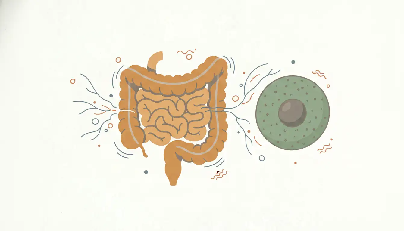
Research in gastroenterology and immunology has established intestinal permeability as a key upstream factor in autoimmune disease development. The intestinal epithelium is a single-cell-layer barrier regulating what crosses from the gut lumen into systemic circulation. Tight junction proteins — occludin, claudin, and zonulin-regulated complexes — control that barrier. When those tight junctions degrade, partially digested food antigens, bacterial lipopolysaccharides, and microbial fragments translocate into circulation and trigger systemic immune activation.
The clinical relevance is direct: you cannot achieve durable remission in autoimmune patients while intestinal permeability remains active. Zonulin is a measurable serum marker for tight junction dysfunction and is part of a rational gut-autoimmunity workup. Stool testing for secretory IgA, the mucosal immune system's first line of defense, gives additional information about whether the gut immune barrier is compromised. These are not exotic tests — they are mechanistically grounded assessments that map directly onto the pathophysiology.
Dr. Kharrazian's clinical training teaches practitioners to treat intestinal permeability not as a downstream symptom but as a primary driver that must be addressed before other autoimmune interventions will hold.
Conducting a Structured Clinical Autoimmune Evaluation
A clinical autoimmune evaluation is not a single lab panel. It is a sequential thought process that moves from symptom pattern recognition to mechanism identification to targeted testing.
The history should focus on three categories: environmental exposures (infections, chemical exposures, dietary history, geographic moves), timeline of symptom onset relative to identifiable triggers, and family history of autoimmune conditions. Autoimmune diseases cluster in families — not always the same disease, but along shared immunological pathways. A patient with Hashimoto's thyroiditis whose mother has rheumatoid arthritis and whose sister has celiac disease is not presenting three separate problems; she is presenting one immunological vulnerability expressed across three tissue types.
Physical examination findings that suggest systemic autoimmune activity include mucosal changes, skin findings consistent with lupus or psoriasis, joint involvement, thyroid enlargement, and neurological soft signs. Questionnaire-based symptom assessments covering multiple organ systems help identify patterns that single-organ specialists may miss because no individual finding is severe enough to trigger a referral.
Laboratory assessment should be layered. Start with tissue-specific autoantibodies based on the clinical picture, not a blanket panel. Add inflammatory markers — high-sensitivity CRP, ESR, ferritin — to assess inflammatory burden. Evaluate gut health markers (zonulin, secretory IgA, calprotectin) to assess the intestinal component. Assess regulatory T cell function indirectly through cytokine patterns where accessible. The goal is not to find every abnormal value but to construct a coherent mechanistic picture of what is driving immune dysregulation in this specific patient.
Why Autoimmune Patients Require Different Clinical Logic
Standard disease management for autoimmune conditions focuses on suppressing the immune response — corticosteroids, biologics, immunosuppressants. That approach reduces symptoms by reducing immune activity broadly. It does not address the mechanisms that initiated and perpetuate the autoimmune process.
Functional medicine autoimmunity training teaches a different clinical logic: identify the environmental triggers sustaining the autoimmune response, restore the barrier functions (intestinal, mucosal, blood-brain barrier) that have been compromised, and support regulatory immune function rather than suppressing immune function globally. This is not a rejection of pharmaceutical management in appropriate cases. It is the addition of a mechanistic layer that standard management does not address.
The distinction matters most for patients with multiple autoimmune diagnoses, patients whose disease continues to progress despite standard treatment, and patients who have positive autoantibodies but have not yet developed diagnosable disease. That last group is where the clinical opportunity is largest — autoantibodies can be detectable years before tissue damage becomes irreversible.
Applying Autoimmune Mechanisms to Complex Cases
The patient presenting with fatigue, joint pain, brain fog, and gastrointestinal symptoms who has been evaluated by multiple specialists and received multiple conflicting diagnoses is not a diagnostic mystery. She is a patient with multiple tissue targets and an unaddressed upstream driver. The mechanism is not mysterious; the clinical evaluation has simply not been designed to see it.
Dr. Kharrazian's coursework at the Kharrazian Institute teaches practitioners to apply autoimmune disease mechanisms systematically — moving from the environmental trigger history through the barrier dysfunction evidence to the tissue-specific antibody pattern — and to build clinical protocols that address each layer. That sequence of thought is what separates a practitioner who manages autoimmune symptoms from one who understands and addresses autoimmune pathophysiology.
The research in functional medicine autoimmunity is advancing faster than most clinical training programs reflect. Practitioners working with chronic, complex autoimmune patients need a continuously evolving clinical education to keep pace with it.
Key Takeaways
- Loss of self-tolerance involves three distinct mechanisms — central tolerance, peripheral tolerance, and regulatory T cell suppression — and failure in any one can initiate autoimmunity given the right environmental context.
- Molecular mimicry, bystander activation, and epitope spreading are the primary mechanisms by which environmental triggers convert genetic susceptibility into active autoimmune pathology.
- Intestinal permeability is a prerequisite for systemic autoimmune activity in most cases and must be directly assessed and addressed for clinical protocols to hold.
- Autoantibodies can be detectable years before diagnosable tissue damage — making early, targeted antibody testing one of the highest-value clinical interventions for at-risk patients.
- A structured autoimmune evaluation sequences from environmental exposure history to barrier function assessment to tissue-specific antibody testing, not from a blanket panel to a diagnosis.
Frequently Asked Questions
Central tolerance eliminates autoreactive T and B cells during development in the thymus and bone marrow. Peripheral tolerance suppresses autoreactive cells that escape into circulation, primarily through regulatory T cells. Autoimmunity can develop when either mechanism is compromised, or when the inflammatory environment overwhelms peripheral suppression.
Molecular mimicry occurs when a foreign antigen shares structural similarity with a self-antigen. The immune response generated against the foreign antigen cross-reacts with the body's own tissue. Gluten-transglutaminase homology and Epstein-Barr virus-myelin cross-reactivity are well-documented examples with direct clinical relevance.
Intestinal permeability allows partially digested food antigens and bacterial fragments to enter systemic circulation, triggering immune activation that can initiate or sustain autoimmune responses. Addressing permeability is a foundational step in functional medicine autoimmunity protocols because other interventions are less effective while the gut barrier remains compromised.
Tissue-specific autoantibodies targeted to the clinical presentation, high-sensitivity CRP and ESR for inflammatory burden, and gut barrier markers including zonulin and secretory IgA form the core of a mechanistic autoimmune workup. The selection should be driven by mechanism, not by reflexive panel ordering.
Epitope spreading is the process by which autoimmunity expands over time as tissue damage exposes hidden self-antigens to the immune system. Clinically, it explains why patients often develop additional autoantibodies as disease progresses and why serial antibody testing over time captures disease trajectory better than a single assessment.
About the Author
Dr. Datis Kharrazian, PhD, DHSc, DC, MS, MMSc, FACN is a Harvard Medical School research fellow and researcher at Massachusetts General Hospital's Department of Neurology specializing in autoimmunity and neuroimmunology. He is an Associate Clinical Professor at Loma Linda University School of Medicine and the author of Why Do I Still Have Thyroid Symptoms When My Lab Tests Are Normal and Why Isn't My Brain Working. He is a Fellow of the American College of Nutrition, a Diplomate of the Board of Nutrition Specialists, a member of the American Association of Immunologists, and a Fellow of the Royal Society of Medicine (UK). The Kharrazian Institute serves more than 5,000 physicians and healthcare providers worldwide.
Practitioner Training in Clinical Autoimmunity
The Kharrazian Institute offers advanced clinical training in autoimmune disease mechanisms, evaluation strategies, and evidence-based protocols for complex autoimmune cases. Coursework is designed for immediate clinical application.