Brain ATP drops, and cognition follows. That single cause-and-effect chain sits at the center of a commonly missed clinical pattern — one that Dr. Datis Kharrazian addresses directly in the neurology coursework at the Kharrazian Institute. The pattern is functional hypoxia: chronic, low-grade oxygen insufficiency that never triggers an emergency but steadily erodes neuronal performance over months or years. Most patients presenting with cognitive fog, attention deficits, and mental fatigue have been evaluated for depression, thyroid dysfunction, and sleep disorders. Oxygen delivery rarely enters the differential.
It should.
Why the Brain Is Uniquely Vulnerable to Reduced Oxygen Availability
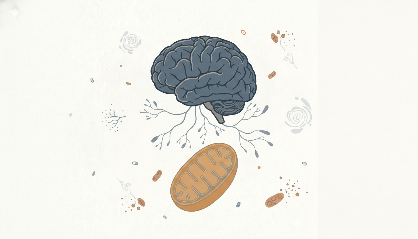
The brain accounts for roughly 20 percent of the body's total oxygen consumption despite representing only about 2 percent of body weight. That disproportionate demand reflects the metabolic cost of maintaining neuronal membrane potentials, synthesizing neurotransmitters, and sustaining synaptic signaling. All of it depends on oxidative phosphorylation — the mitochondrial process by which oxygen is converted into ATP.
When oxygen availability drops even modestly, mitochondrial ATP production becomes insufficient to sustain normal neuronal activity. The brain has no meaningful glycogen reserve to compensate. Neurons begin operating below their functional threshold, and the clinical result is exactly what practitioners observe: reduced processing speed, poor working memory, difficulty sustaining attention, and a generalized sense of cognitive heaviness that patients often describe as their brain "not turning on."
This is not anoxia. It is not a stroke. It is a functional energy deficit — chronic, subclinical, and frequently misattributed to mood disorders or aging.
How Hypoxia Initiates Neuroinflammation
Oxygen insufficiency does more than reduce ATP output. It activates hypoxia-inducible factor pathways that upregulate pro-inflammatory gene expression within the central nervous system. Research in neuroimmunology demonstrates that sustained hypoxic signaling drives microglial activation — and once microglia shift to a primed or reactive state, they release cytokines that amplify neuronal stress independent of the original oxygen deficit.
The clinical implication is a self-reinforcing loop. Hypoxia produces neuroinflammation. Neuroinflammation impairs mitochondrial function. Impaired mitochondria reduce the neuron's capacity to tolerate any further metabolic stress, including subsequent hypoxic episodes. Practitioners treating cognitive decline who address inflammation without identifying the hypoxic driver are managing a downstream consequence while the upstream cause persists.
Dr. Kharrazian's clinical training emphasizes that neuroinflammation rarely has a single cause, and that identifying oxygen delivery as a contributing variable — particularly in patients with respiratory dysfunction, cardiovascular compromise, or anemia — is a necessary step in building an accurate clinical picture.
The Role of Glial Cells in Hypoxia-Driven Cognitive Decline
Neurons are not the only cells affected. Astrocytes and microglia, collectively the glial cell populations that regulate neuronal homeostasis, are highly sensitive to hypoxic conditions. Under normal oxygen levels, astrocytes supply neurons with metabolic substrates and regulate the extracellular environment. When hypoxia primes these cells toward inflammatory signaling, that supportive function is compromised.
Primed microglia, in particular, lower the activation threshold for subsequent inflammatory responses. A patient whose microglia are already sensitized by chronic hypoxia will have an exaggerated neuroinflammatory response to otherwise tolerable stressors — a mild infection, a dietary insult, a poor night of sleep. What would be a transient event in a neurologically resilient patient becomes a multi-day cognitive crash in a patient with a primed glial population. This is why some patients report that their cognitive symptoms fluctuate dramatically in response to what appear to be minor provocations.
Recognizing Functional Hypoxia in Patients With Cognitive Complaints
Functional hypoxia does not always present with the classic signs of respiratory distress. Patients are not necessarily dyspneic. They breathe. They have acceptable oxygen saturation on pulse oximetry at rest. The insufficiency is functional — meaning it becomes relevant under metabolic demand, during sustained cognitive effort, or in the context of impaired oxygen delivery mechanisms that never reach the threshold of a formal diagnosis.
The symptom cluster to recognize includes:
- Cognitive fog that worsens with exertion or sustained mental tasks
- Reduced mental endurance — difficulty thinking clearly in the afternoon relative to the morning
- Fatigue disproportionate to activity level
- Slow information processing and word retrieval difficulties
- Mood instability that worsens in tandem with cognitive symptoms
These are not psychiatric symptoms. They are neuroenergetic symptoms, and they map directly to ATP-insufficient neuronal function. Chronic respiratory conditions — sleep apnea, poorly managed asthma, chronic obstructive patterns — are among the most common structural contributors. Anemia, reduced cardiac output, and poor peripheral vascular tone can produce the same picture through different mechanisms. The common denominator is oxygen reaching the brain in quantities insufficient to sustain its metabolic load.
What Systemic Physiological Dysfunction Tells You About Brain Oxygenation
Oxygen delivery to the brain is downstream of multiple systems: pulmonary gas exchange, hemoglobin concentration and function, cardiac output, cerebrovascular tone, and mitochondrial utilization efficiency. A deficiency anywhere in that chain can produce functional hypoxia at the neuronal level.
Practitioners trained in the KI approach to brain-based conditions learn to evaluate each link. Cardiovascular compromise reduces cerebral perfusion pressure. Iron-deficiency anemia reduces oxygen-carrying capacity even when saturation appears normal. Mitochondrial dysfunction, whether from nutrient deficiencies, toxin exposure, or chronic inflammation, impairs the neuron's ability to extract and utilize available oxygen effectively — a state that can produce hypoxic physiology even when delivery is technically adequate.
This last point is clinically significant. A patient can have normal hemoglobin, normal oxygen saturation, and normal cardiac output and still present with functional neuronal hypoxia if mitochondrial utilization is compromised. The research synthesized in Dr. Kharrazian's coursework addresses this distinction and teaches practitioners to evaluate systemic physiological dysfunction as a whole rather than ruling out hypoxia based on a single normal value.
Clinical Strategies for Addressing Functional Hypoxia and Cognitive Decline
Identifying functional hypoxia as a contributor to cognitive decline shifts the clinical strategy considerably. The goal becomes restoring adequate oxygen delivery and mitochondrial utilization efficiency, not suppressing symptoms.
Cardiovascular fitness is one of the most well-established variables in cerebral oxygenation. Research in exercise physiology and neurology consistently demonstrates that aerobic conditioning improves cerebrovascular tone, increases cerebral blood flow, and upregulates mitochondrial biogenesis. For patients who are deconditioned, even low-intensity, progressive aerobic activity produces measurable improvements in cognitive function — not through psychological mechanisms, but through direct improvements in brain oxygen delivery.
Addressing underlying chronic respiratory conditions is equally direct. Untreated obstructive sleep apnea produces recurrent nocturnal hypoxic events that prime microglial populations over time. Research on sleep apnea and cognitive decline shows a dose-response relationship: the longer the condition goes unaddressed, the more significant the neuroinflammatory burden. Effective management of sleep-disordered breathing is, by extension, a neurological intervention.
Nutritional variables that support mitochondrial function — B vitamins (particularly B1, B2, and B3 as cofactors in the electron transport chain), CoQ10, iron, and magnesium — are relevant to the oxygen utilization side of the equation. These are not supplements for cognitive enhancement in a healthy population; they are corrective interventions when deficiencies are impairing the machinery that converts oxygen into neuronal energy. The clinical application is personalized, based on laboratory evaluation and a thorough intake of the patient's metabolic history.
Dr. Kharrazian's clinical training integrates these variables into a coherent sequence of thought: identify the mechanism, locate the breakdown in the oxygen delivery chain, and build a protocol that addresses the specific deficiency. That sequence is what distinguishes targeted intervention from generic cognitive support.
Key Takeaways
- The brain's disproportionate oxygen demand makes it acutely sensitive to even modest reductions in oxygen availability, producing ATP deficits that manifest as cognitive fog, reduced mental endurance, and attentional impairment.
- Hypoxia activates neuroinflammatory pathways and primes glial cells, creating a cycle that perpetuates neuronal dysfunction beyond the initial oxygen deficit.
- Functional hypoxia can exist with normal pulse oximetry if mitochondrial utilization is impaired — normal saturation does not rule out neuronal oxygen insufficiency.
- Chronic respiratory conditions, anemia, reduced cardiac output, and mitochondrial dysfunction are all mechanisms capable of producing hypoxia-driven cognitive decline.
- Effective clinical strategies target the specific point of failure in the oxygen delivery chain, including aerobic conditioning, management of sleep-disordered breathing, and correction of nutritional deficiencies that impair mitochondrial function.
Frequently Asked Questions
Clinical hypoxia is a measurable, often acute drop in blood oxygen saturation. Functional hypoxia refers to chronic, subclinical oxygen insufficiency at the cellular level — particularly in neurons — where delivery or utilization is inadequate to sustain metabolic demand without producing emergency-level symptoms or abnormal pulse oximetry readings.
When brain ATP drops, neurotransmitter synthesis is impaired along with all other energy-dependent neuronal processes. Dopamine and serotonin production both require adequate mitochondrial energy. Patients with hypoxia-driven cognitive decline frequently present with concurrent mood instability that resolves when oxygenation and mitochondrial function are addressed.
Obstructive sleep apnea produces repeated nocturnal hypoxic events that activate and prime microglial populations over time. Research demonstrates a dose-response relationship between untreated sleep apnea duration and neuroinflammatory burden, making chronic respiratory conditions a direct structural contributor to progressive cognitive decline.
Mitochondrial dysfunction impairs oxygen utilization efficiency at the cellular level. Even with adequate oxygen delivery, neurons unable to convert oxygen into ATP effectively will behave as though hypoxic. B vitamin deficiencies, CoQ10 insufficiency, and chronic inflammatory states are common contributors to this utilization failure.
The Kharrazian Institute's brain-based clinical coursework teaches practitioners to evaluate the full oxygen delivery chain — from pulmonary function and hemoglobin status through mitochondrial utilization — and to build personalized protocols for patients presenting with cognitive decline and systemic physiological dysfunction.
About the Author
Dr. Datis Kharrazian, PhD, DHSc, DC, MS, MMSc, FACN is a Harvard Medical School research fellow and researcher at Massachusetts General Hospital Department of Neurology specializing in autoimmunity and neuroimmunology. He serves as Associate Clinical Professor at Loma Linda University School of Medicine and is the author of Why Do I Still Have Thyroid Symptoms When My Lab Tests Are Normal and Why Isn't My Brain Working. Dr. Kharrazian is a Fellow of the American College of Nutrition, Diplomate of the Board of Nutrition Specialists, member of the American Association of Immunologists, and Fellow of the Royal Society of Medicine (UK). The Kharrazian Institute serves more than 5,000 physicians and healthcare providers worldwide.
Clinical Training in Brain-Based Conditions
The Kharrazian Institute offers advanced clinical coursework on neurological dysfunction, neuroinflammation, and cognitive decline. Practitioners looking to apply evidence-based strategies for identifying and addressing functional hypoxia and its downstream neurological effects can explore current course offerings at the Kharrazian Institute.